Sporoschisma aquaticum Z.L. Luo, H.Y. Su&K.D.Hyde, sp. nov.,
Index Fungorum number: IF 818333; MycoBank number: MB 818333; Facesoffungi number: FoF 02374
Etymology – In reference to the aquatic habitats of this fungus.
Holotype – DLU 628
Saprobic on submerged decaying wood. Sexual morph: Undetermined. Asexual morph: Colonies on the substratum superficial, effuse, gregarious, hairy, pale black. Mycelium immersed, composed of pale to dark brown hyphae. Setae scattered, capitate, with hyaline, mucilaginous substances at the swollen apex, smooth-walled, pale brown, becoming paler towards the apex, straight or flexuous, septate, 127 − 141 × 4 – 6 μm (x = 134 × 5 μm, n = 10). Conidiophores macronematous, mononematous, smooth, black, straight or slightly flexuous, solitary, each composed of a bulbous base, a cylindrical stipe and a swollen venter, erect, 183–207 μm long, 7–9 μm wide below venter and 9.5–12.5 μm wide above, 15–20 μm wide at venter. Conidiogenous cells monophialidic, integrated, terminal, determinate, brown. Conidia catenate, cylindrical, 26–32 × 9–10 μm (x = 29 × 9.5 μm, n=25), 3-septate, verruculose, brown.
Material examined – CHINA, Yunnan Province, saprobic on decaying wood submerged in the Lancang River, April 2014, Z.L. Luo, HL 23–8–2 (DLU 628, holotype), ex-type living culture, DLUCC 0628.
Notes – Sporoschisma aquaticum was collected from northwestern Yunnan Province on submerged wood in Lancang River. Sporoschisma aquaticum is morphologically similar to S. palauense and S. juvenile, but S. aquaticum differs from S. palauense in having shorter setae (127 − 141 μm vs. 115−215 μm), verruculose, and smaller conidia (26–32 × 9–10 μm vs. 26–58 × 10–14 μm) whereas the conidia of S. palauense have a conspicuous, spherical guttule in almost all cells. S. aquaticum differs from S. juvenile in having shorter conidiophores (183–207 μm vs. up to 280 μm), and smaller conidia (26–32 × 9–10 μm vs. 34–44 × 10.5–14.5 μm).
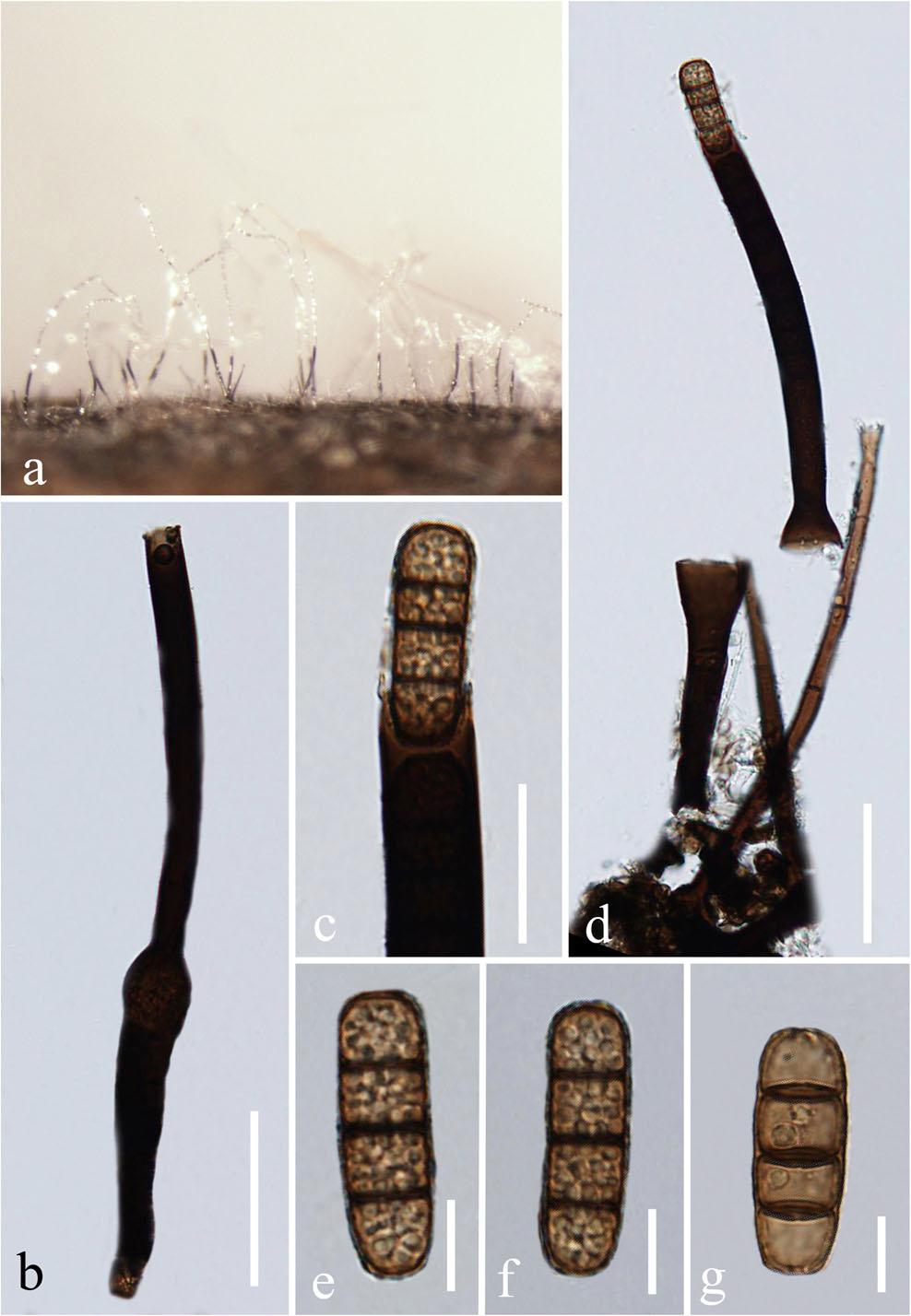
Fig 1. Sporoschisma aquaticum (DLU 628, holotype) a Colonies on wood. b Conidiophore. c Portion of phialide producing conidia. d Conidiophore with setae. e − g Conidia. Scale bars: b, d = 50 μm, c = 20 μm, e–g = 10 μm.