Sporoschisma hemipsilum (Berk.&Broome) Zelski, A.N. Mill., & Shearer, IMA Fungus 5(2): 433 (2014).
Basionym: Sphaeria hemipsila Berk. & Broome, Bot. J. Linn. Soc. 14: 126 (1873).
Synonyms: Lasiosphaeria hemipsila (Berk. & Broome) Sacc., Syll. Fung. 2: 198 (1883).
Chaetosphaeria hemipsila (Berk. & Broome) Petch., Ann. Roy. Bot. Gard. Peradenija 6: 336 (1917).
Melanochaeta hemipsila (Berk. & Broome) E. Müll. et al., Revue Mycol. 33: 377 (1969).
Chaetosphaeria coelestina Höhn., Sitzungsber. Akad. Wiss. Wein, Math.-Naturwiss. Kl, 1 Abt. 118: 324 (1909).
Sporoschisma saccardoi E.W. Mason&S. Hughes, Mycol Pap. 31: 20 (1949).
Index Fungorum number: IF 816542; Facesoffungi number: FoF 02376
Saprobic on submerged, decaying wood. Sexual morph: See Goh et al., Mycol Res. 101(II): 1303 (1997). Asexual morph: Colonies on the substratum superficial, effuse, hairy, gregarious, pale black to black. Mycelium immersed, composed of pale to dark brown hyphae. Setae scattered or in groups mixed with conidiophores, capitate, usually with hyaline, mucilaginous substances at the swollen apex, smooth-walled, pale brown, becoming paler towards the apex, straight or flexuous, 1 − 3 septate, 165.5− 185.5 × 5–6 μm (x = 175.5 × 5.5 μm, n = 10). Conidiophores macronematous, mononematous, smooth, dark brown to black, straight or slightly flexuous, solitary or in small groups, each composed of a bulbous base, a cylindrical stipe and a swollen venter with a long cylindrical neck, erect, 238–258 μm long, 8–11 μm wide below venter and 15–19 μm wide above, 19–21 μm wide at venter. Conidiogenous cells monophialidic, integrated, terminal, determinate, brown, lageniform, with serrate, flared margin at free end. Conidia formed in chains, cylindrical to doliiform, 42–47 × 12.5–13.5 μm (x = 42×13 μm, n = 30), 5-septate, with conspicuously darkened septa, occasionally constricted at septa, center cells brown, end cells paler coloured and shorter than the central four cells.
Material examined – CHINA, Yunnan Province, saprobic on decaying wood submerged in Nujiang River, May 2015, S.M. Tan, N 6–8–1 (HKAS 92767), living culture, KUMCC 15–0489, MFLUCC; HL 5-12-1 (HKAS 92843); 2LQXM 18-2; N4-2-1.
Notes – This fungus was recovered from a variety of habitats with a range of environmental conditions. Cai et al. (2002) and Luo et al. (2004) have recorded this species from Lake Fuxian and Dianchi in Yunnan Province, China. In this study, we provided new descriptions and illustrations of Sporoschisma hemipsilum.
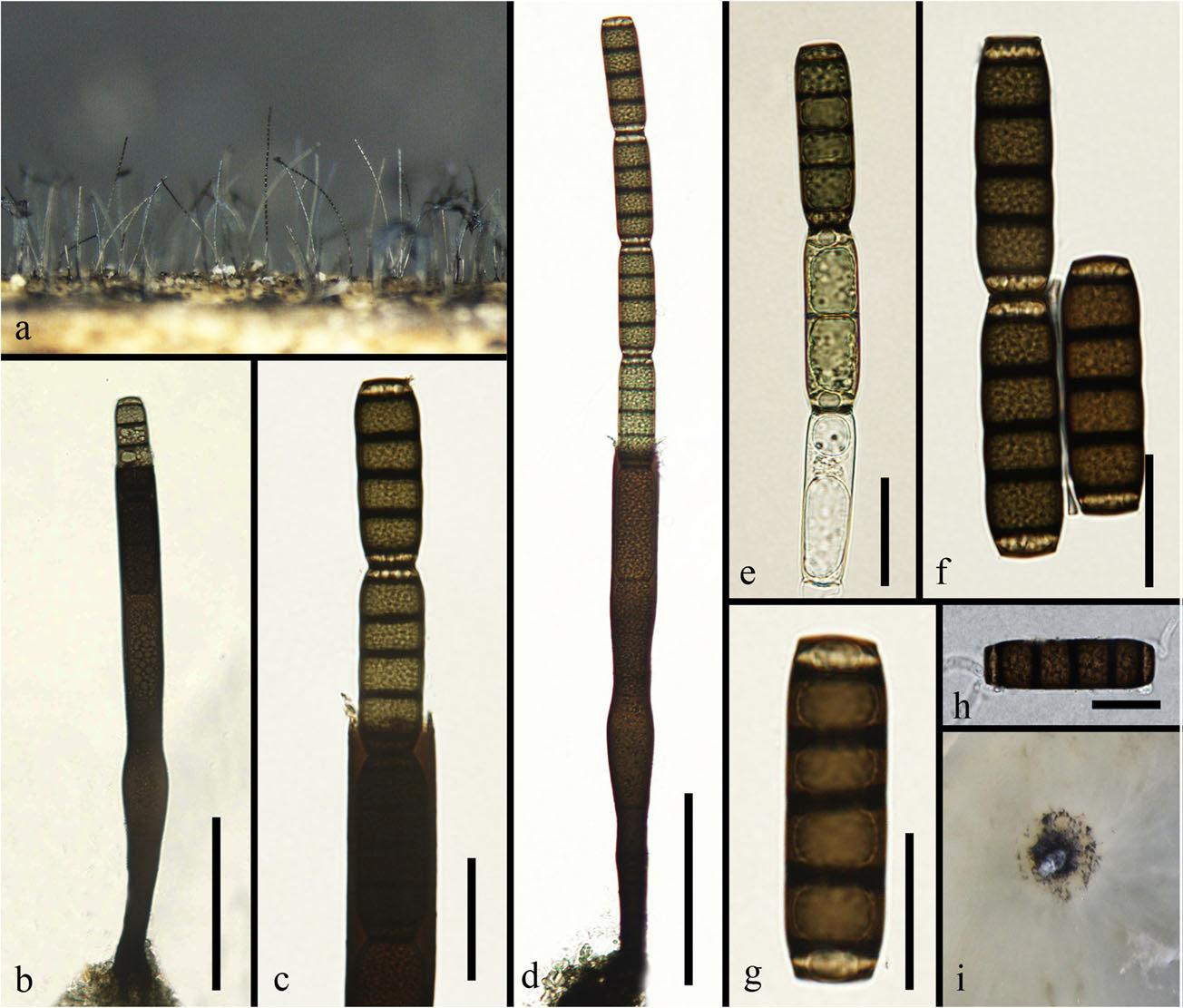
Fig 1. Sporoschisma hemipsilum (HKAS 92767) a Colonies on wood. b Conidiophore. c Portion of phialide producing conidia. d Conidiophore and seta. e–g Conidia. h Germinated conidium. i Culture on PDA. Scale bars: d = 100 μm, b = 80 μm, c = 30 μm, e, f = 30 μm, g, h = 20 μm.