Phaeosphaeria oryzae I. Miyake, Bot. Mag., Tokyo 23: 93 (1909)
= Phaeoseptoria oryzae I. Miyake (1910).
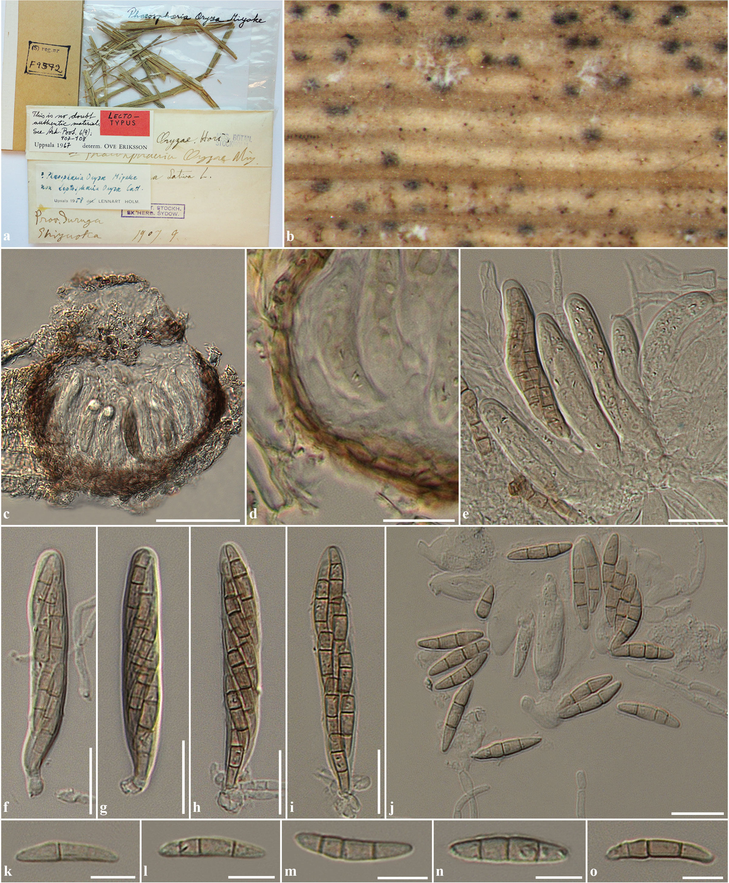
Saprobic on leaves of Oryza sativa. Ascomata 70–130 µm high, 90–150 µm diam., solitary, sometimes clustered, immersed, visible as slightly raised, small, black dots on the host surface, uniloculate, globose to subglobose, glabrous, brown to dark brown, ostiole central, with minute papilla. Peridium 5–10 µm wide, of equal thickness, composed of 2–3 layers of thin-walled, brown to dark brown, flattened pseudoparenchymatous cells, arranged in textura angularis. Hamathecium composed of sparse, 2.5–4 µm wide, filiform, broad cellular pseudoparaphyses, distinctly constricted at the septa, rarely anastomosing at the apex. Asci (50–)55–70(–80) × 10–13.5(–15) µm ( = 65.4 × 11.9 μm, n = 25), 8-spored, bitunicate, fissitunicate, broadly cylindrical, short pedicellate or subsessile, apically rounded with indistinct ocular chamber. Ascospores (22–)25–27(–29) × 5–6 µm ( = 26 × 5.2 μm, n = 30), overlapping 2-seriate, phragmosporous, narrowly fusiform, brown, mostly 3-septate, rarely 1–2 septa, indistinctly constricted at the septa, slightly curved, smooth-walled. Asexual state: Phaeoseptoria.
Material examined: JAPAN, Shizuoka, Suruya, on Oryza sativa L. (Poaceae), September 1907 (S-F9572, lectotype); EL SALVADOR, intercepted Washington Dc #26812, on Oryza sativa, 10 March 1952, F.P. Hubert (BPI 622541); JAPAN, intercepted San Diego California #4872, on Oryza sativa L., 27 December 1957, E.B. Francis (BPI 622542); JAPAN, intercepted San Francisco California #17349, 17352, 17367, on Oryza sativa, 13 December 1940, F.J. Phelan (BPI 622544); NICARAGUA, Monagua, on living leaves of Oryza sativa, 4 November 1954, C. Pineda 37, (BPI 622545); THAILAND, Chiang Mai Province, Mae Wang District, Mae Sapok Royal Project, on dead branches of Etlingera sp. (Zingiberaceae), 5 October 2010, R. Phookamsak RP0086 (MFLU 11-0206), living culture = MFLUCC 11-0170.
Fig. 1 Phaeosphaeria oryzae (S-F9572, lectotype) a Herbarium label and specimens. b Ascomata on host surface. c Section through ascoma. d Section through peridium. e Asci with sparse pseudoparaphyses. f-i Asci. j-o Ascospores. Scale bars: c = 50 µm, e, f, g, h, i, j = 20 µm, d, k, l, m, n, o = 10 µm.