Ophiosphaerella agrostidis Dern. et al.
Pathogenic or saprobic on monocotyledonous hosts. Sexual state: Ascomata 275–355 µm high (including papilla), 175–310 µm diam., scattered, solitary, semi immersed or erumpent through host tissue with papilla, visible as raised, small black dots on host surface, uniloculate, globose to subglobose, glabrous, dark brown to black, ostiole central, oblique, papilla, carbonaceous. Peridium 15–25 µm wide, thin-walled, of equal thickness, composed of several layers of brown to dark brown, pseudoparenchymatous cells, arranged in a textura angularis to textura globulosa. Hamathecium composed of numerous, 1.5–2.5 µm wide, filamentous, broad cellular pseudoparaphyses, with distinct septa, embedded in mucilaginous matrix, anastomosing at the apex. Asci (97–)100–135 × 7–9(–11) µm ( = 111.5 × 8.5 μm, n = 25), 8-spored, bitunicate, fissitunicate, cylindrical to cylindric-clavate, short pedicellate, apically rounded, with a well-developed ocular chamber. Ascospores (90–)98–120(–130) × 2–2.5 µm ( = 110.3× 2.3 μm, n = 30), fasciculate, parallel or spiral, scolecosporous, filiform, narrowing towards ends, pale brown to brown, 15-septate, not constricted at the septa, smooth-walled. Asexual state: Unknown.
Material examined: THAILAND, Phrae, Rongkwang District, Maejo University Phrae Campus, on dead culms of grass, 20 August 2010, R. Phookamsak (MFLU 11-0188), living culture = MFLUCC 11-0152; Chiang Rai, Muang District, Ta Sai, on dead culm of grass, 30 June 2011, R. Phookamsak (MFLU 11-0246), living culture = MFLUCC 12-0007.
Notes: The scolecosporous specimens were collected from dead culms of grass in Thailand. These collections are morphologically similar to Ophiosphaerella agrostidis, Op. graminicola, and Op. herpotricha with these species sharing similar ascomata, asci and ascospores, including septation of ascospores. The Thailand collections are most similar to Op. agrostidis according to the size of the ascomata, asci and ascospores, and also the septation of ascospores. However, Op. agrostidis has been reported as a pathogen on grass while the collections in the present study were saprobes. Therefore these collections are named as Op. agrostidis based on the morphological characters and these can be treated as authentic specimens until epitypification. The phylogenetic tree shows that two isolates of Op. agrostidis form a single clade in Phaeosphaeriaceae (Table 1).
Table 1. Synopsis of Ophiosphaerella species discussed in this study

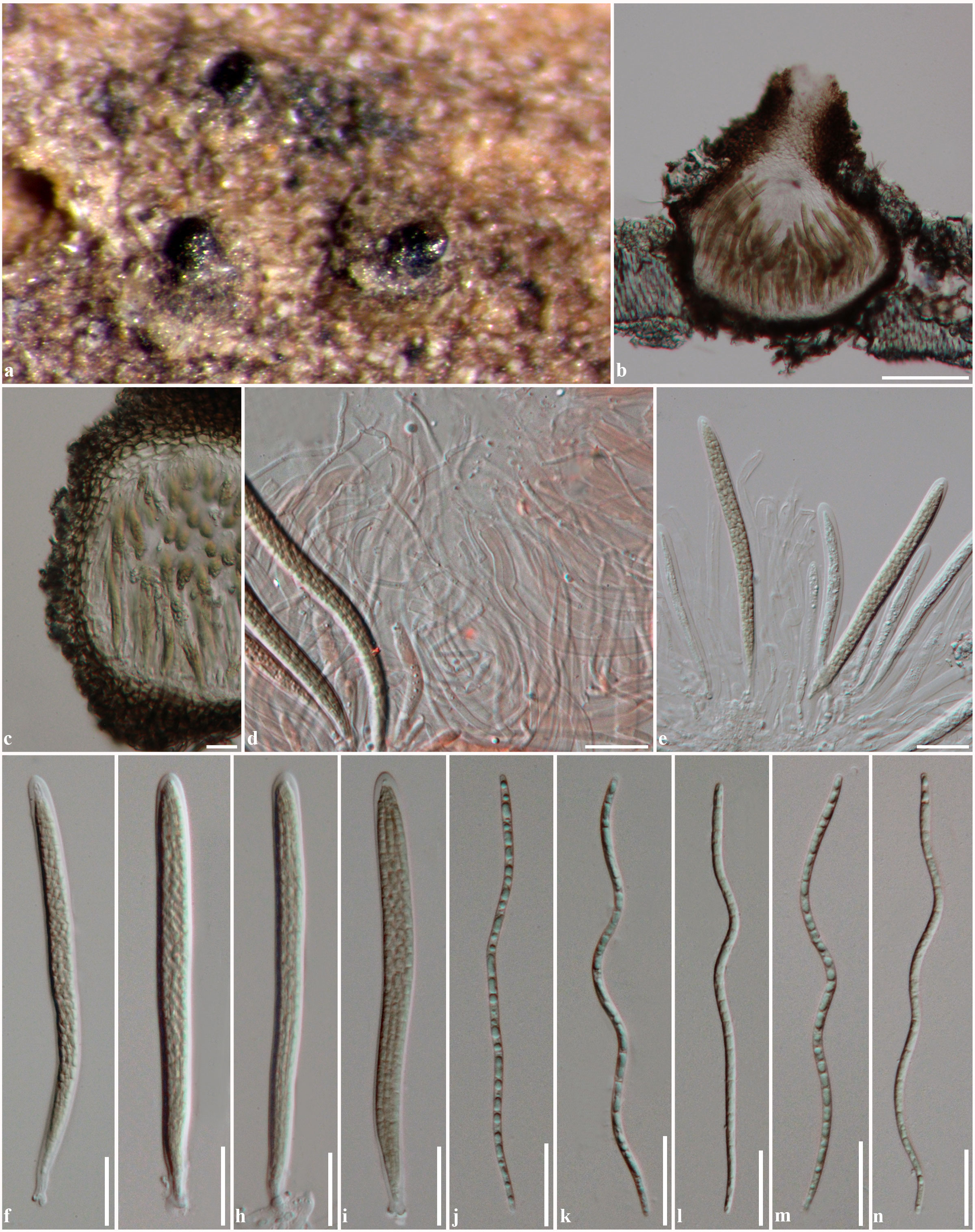
Fig. 1 Ophiosphaerella agrostidis (MFLU 11-0188). a Ascomata on host surface. b Section through ascoma c Section through peridium. d Cellular pseudoparaphyses stained in congo red. e-i Asci. j-n Ascospores. Scale bars: b = 100 µm, c–n = 20 µm.