Phaeosphaeria chiangraina Phookamsak & K.D. Hyde, sp. nov., Index Fungorum number: IF550736
Etymology: Referring to Province in Thailand, in which the fungus was found.
Holotypus: MFLU 12-2472
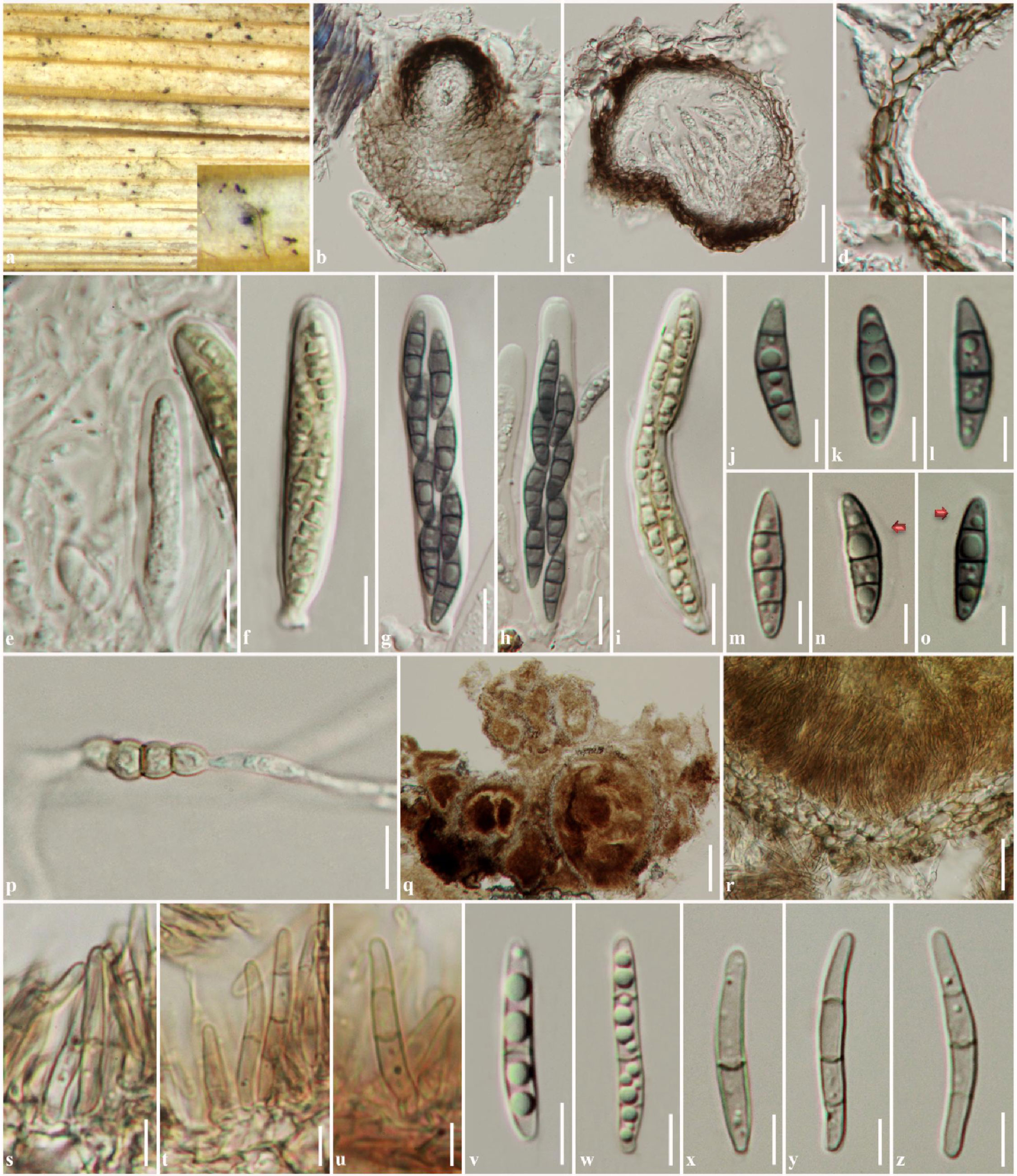
Saprobic on leaves of Oryza sativa. Sexual state: Ascomata 120–180 µm high, 130–180 µm diam., scattered, immersed, visible as slightly raised, small, black dots on the host surface, uniloculate, globose to subglobose or irregular in shape, glabrous, brown to dark brown, ostiole central with minute papilla. Peridium 7–16 µm wide, thin-walled, of equal thickness, composed of 1–3 layers of flattened, brown to dark brown, elongated, pseudoparenchymatous cells, arranged in textura angularis. Hamathecium composed of numerous, 1.5–3 µm wide, filiform, broad cellular pseudoparaphyses, with small guttules, indistinctly constricted at the septa, embedded in mucilaginous matrix, rarely anastomosing at the apex. Asci (44–)47–60(–65) × 8–9(–10) µm ( = 52.6 × 8.9 μm, n = 25), 8-spored, bitunicate, fissitunicate, broadly cylindrical, short pedicellate or subsessile, apically rounded with ocular chamber. Ascospores (13.5–)15–17(–18) × 3.5–4.5 µm ( = 16.4 × 4.1 μm, n = 30), overlapping 2-seriate, phragmosporous, fusiform with obtuse ends, pale brown when young, becoming yellowish-brown to brown at maturity, dark brown when stained in 5% of KOH, 3-septate, not constricted at the septa, slightly curved, smooth-walled with small to large guttules in each cell, surrounded by a distinct mucilaginous sheath. Asexual state: produced on media agar after 8 weeks. Conidiomata 100–305 µm high, 120–290 µm diam., pycnidial, scattered to clustered, immersed, with dark brown to black stromatic tissues embedded in the media agar, uni- to multi-loculate, globose to irregular, ostioles not observed, brown to dark brown. Conidiomata walls 13–35 µm wide, composed of several layers of brown, pseudoparenchymatous cells, arranged in a textura angularis, covered by vegetative hyphae. Conidiophores reduced to conidiogenous cells. Conidiogenous cells (2–)3–6 × (2–)4–7 µm ( = 4.4 × 4.3 μm, n = 20), holoblastic, phialidic, ampulliform or flattened, hyaline to pale brown, difficult to distinguish from the pycnidial wall. Conidia (15–)17–21.5(–26) × 2–3 µm ( = 19.4 × 2.5 μm, n = 30), solitary, cylindrical, pale brown to brown, 1–3-septate, slightly curved, obtuse at the apex, truncate at the base, not constricted at the septa, smooth-walled with guttules.
Culture characters: Colonies on PDA 77–80 mm diam. after 4 weeks at 25–30 ◦C, white to pale yellowish at the edge, pale yellowish to grey in the centre, forming different colour sectors of grey to dark grey; reverse pale yellowish at the edges, becoming yellowish-brown to brown at the centre, slightly radiating outwardly, forming dark sectors; medium dense, forming black stromatic tissues immersed in media after 8 weeks, circular to irregular, flattened to slightly raised, dull with entire edge, floccose to fairly fluffy, slightly radiating at the lower part, not pigmented.
Material examined: THAILAND, Chiang Rai Province, Muang District, Nanglae Village, on dead leaves of Oryza sativa, 20 January 2013, R. Phookamsak RP0131 (MFLU 12-2472, holotype), ex-type living culture = MFLUCC 13-0231 = MUCL.
Notes: Phaeosphaeria chiangraina was collected on Oryza in Thailand. This is the same host of the generic type of Phaeosphaeria, P. oryzae. A morphological comparison based on species described in Shoemaker and Babcock (1989b) shows that Ph. chiangraina is similar to Phaeosphaeria avenaria, Ph. eustoma, Ph. oryzae and Ph. tritici. Phaeosphaeria chiangraina differs from Ph. oryzae in the size of ascomata, asci and ascospores. Phaeosphaeria chiangraina has smaller ascospores (13.5–18 × 3.5–4.5 μm) than Ph. oryzae (15–23 × 4–5 μm). Phaeosphaeria tritici has similar sized ascospores to Ph. chiangraina; however, Ph. tritici has a larger asci and smaller ascomata. Additionally, Phaeosphaeria tritici ascospores are enlarged at the second cell and slightly constricted at the first septa and produce a phoma-like asexual state, as while Ph. chiangraina lacks an enlarged cell, is not constricted at the septa and forms a septoria-like asexual state.
Multigene phylogenetic analysis using MP shows that Phaeosphaeria chiangraina can be distinguished from Phaeosphaeria avenaria and Ph. eustoma, and forms a strongly supported clade with Ph. thysanolaenicola and Ph. oryzae (CBS 110110) (Fig. 1). However, Ph. chiangraina morphologically differs from Ph. thysanolaenicola and can be separated from the epitype of Ph. oryzae due to its smooth-walled ascospores with a distinct sheath and based on phylogenetic evidence.
Fig. 1 Phaeosphaeria chiangraina (MFLU 12-2472, holotype). a Ascomata of Phaeosphaeria oryzae on host surface. b Ascomata visualized under compound microscope. c Section through ascoma. d Section through peridium. e Asci with rare pseudoparaphyses. f, i Asci. g, h Asci stained in 5% KOH. j- o Ascospores stained in 5% of KOH. m, n Ascospores. p Ascospore germination on WA. q Section through conidioma. r Section through conidioma wall. s–u Conidiogenous cells. v–z Conidia. Scale bars: q = 100 µm, b, c = 50 µm, d, r = 20 µm, e, f, g, h, i, p = 10 µm, j, k, l, m, n, o, s, t, u, v, w, x, y, z = 5 µm.