Paraphaeosphaeria michotii (Westend.) O.E. Erikss., Cryptogams of the Himalayas 6: 405 (1967)
Basionym: Sphaeria michotii Westend., Bull. Acad. R. Sci. Belg., Cl. Sci., sér. 2 7(5): 87 (1859).
Index Fungorum number: IF 335615; MycoBank number: MB 335615; Facesoffungi number: FoF 00058
Saprobic on dead leaves of Poaceae. Sexual state: Ascomata 130–200 × 150–250 µm (x̅ = 150 × 320 µm, n = 10) small to medium-sized, immersed to semi immersed, depressed globose, ostiolate. Ostiole papillate, black, smooth, with beak and without periphyses. Peridium 10–17 μm (x̅ = 14 µm, n = 20) wide, usually with 3–5 layers, composed of cells textura prismatica. Hamathecium of dense 2–3 μm (x̅ = 2 µm, n = 20) filamentous, hyaline, septate, broad, pseudoparaphyses. Asci 60–85 × 12–28 μm (x̅ = 77 × 20 µm, n = 20), 8-spored, bitunicate, fissitunicate, cylindrical with a short, broad pedicel with minute ocular chamber. Ascospores 15–30 × 4–7 μm (x̅ = 24 × 5 μm, n = 40), uniseriate or partially overlapping, 2-septate, broadly elliptical, yellowish-brown, with small guttules, smooth, with a thick uniform sheath. Asexual state: not observed in the culture.
Material examined – ITALY, Forlì-Cesena, Montevescovo, on dead leaves of Poaceae, 3 February 2012, E. Camporesi (MFLU 12-2210, epitype), ex-type living culture = MFLUCC 13-0349.
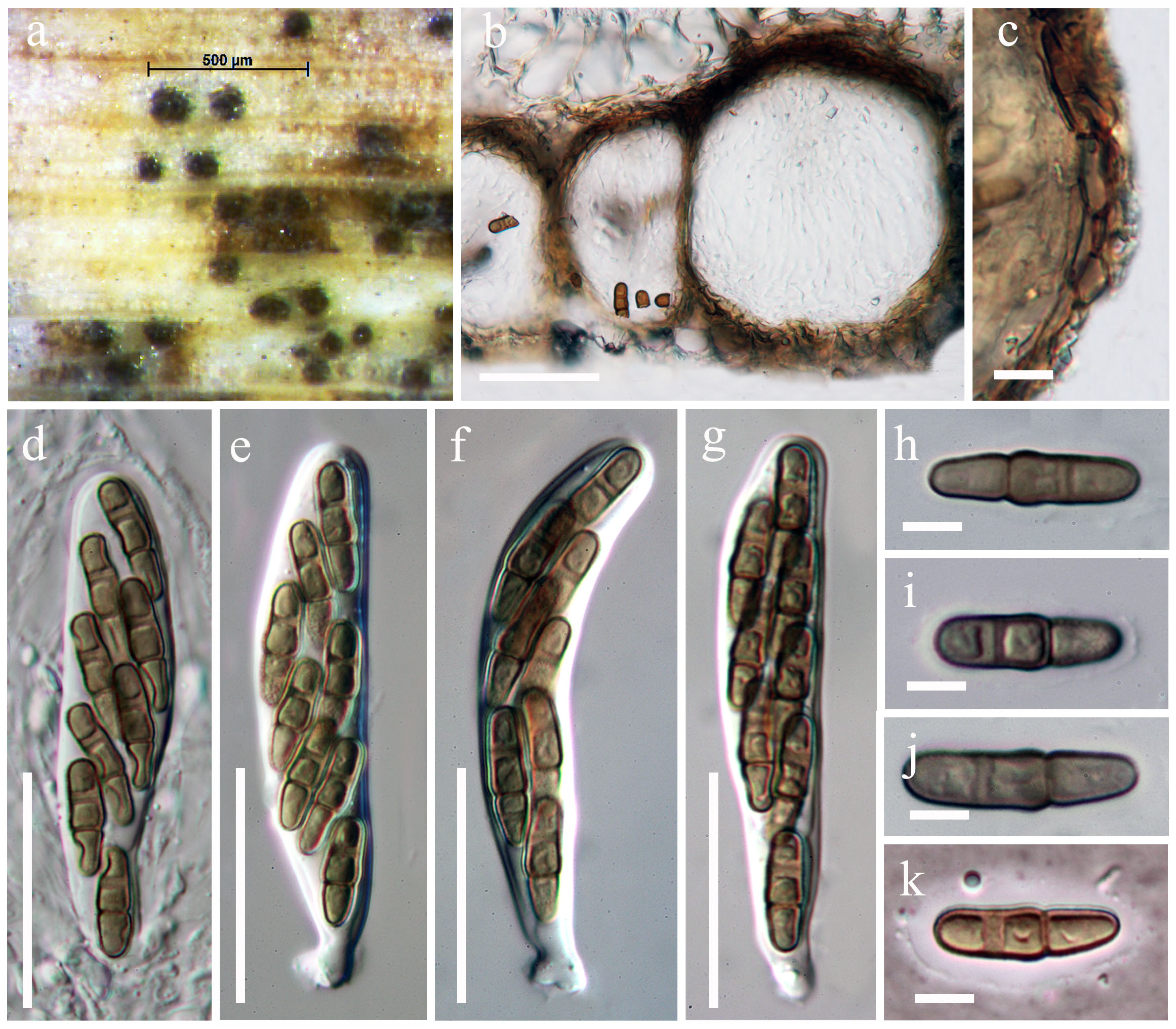
Fig. 1 Paraphaeosphaeria michotii (MFLU 12-2210, epitype). a Ascomata on host substrate. b Section of ascoma. c Close up of the peridium. d Ascus surrounded by cellular pseudoparaphyses. e–g Asci with short, broad pedicel bearing 8 spores. h–j Mature ascospores with thin uniform sheath. k Ascospores mounted in Indian ink. Scale bars: b = 100 μm, c = 50 μm, d = 20 μm, f–h = 60 μm, i–k = 10 μm.
Notes – Paraphaeosphaeria has been separated from Leptosphaeria (Eriksson 1967), and it is quite comparable with Phaeosphaeria. Paraphaeosphaeria can be distinguished from Phaeosphaeria by its ascospores. Ascospores of Paraphaeosphaeria michotii have two septa, and they are biseriate, straight, subcylindrical with broadly rounded ends, rather dark brown and punctate. The primary septum is laid down closer to the distal end than to the proximal, and the larger, proximal hemispore is divided by one transverse septum. There are more septa in the proximal hemispore of other species such as Paraph. castagnei (Durieu & Mont.) O.E. Erikss., Paraph. obtusispora (Speg.) O.E. Erikss. and Paraph. vectis (Berk. & Broome) Hedjar. Asexual characters can also distinguish Paraphaeosphaeria and Phaeosphaeria. Paraphaeosphaeria produces Coniothyrium-like asexual states, but Phaeosphaeria has Hendersonia–Phaeoseptoria asexual states (Eriksson 1967). Câmara et al. (2001) provided descriptions of sexual and asexual morphs of Paraph. michotii and Paraph. pilleata, while other species treated under Paraphaeosphaeria were transferred later to Neophaeosphaeria and Phaeosphaeriopsis (Câmara et al. 2003). None of the amerosporic coniothyrium-like species associated with these sexual morphs has been assigned with a formal name. Shoemaker and Babcock (1985) redescribed some Canadian and extralimital species and excluded Paraph. longispora (Wegelin) Crivelli and Paraph. oblongata (Niessl) Crivelli from Paraphaeosphaeria based on their longitudinal septa as well as beak-like papilla and wall structures. Molecular phylogenetic results based on a multi-gene study indicate that Paraphaeosphaeria should belong to Montagnulaceae (Zhang et al. 2009a). Recently, Ariyawansa et al. (2014d) epitypified Paraph. michotii from a fresh collection and thus the placement of the Paraphaeosphaeria in Montagnulaceae is confirmed. In our phylogenetic analysis, Paraphaeosphaeria forms a well-supported clade sister to Didymosphaeria, thus Paraphaeosphaeria is treated as a separate genus in the Didymosphaeriaceae.