Ophiosphaerella graminicola Speg., Anal. Mus. nac. B. Aires, Ser. 3 12: 401 (1909)
Pathogenic or saprobic on monocotyledons. Sexual state: Ascomata 230–300 µm high, 170–230 µm diam., scattered, solitary, immersed to semi immersed or erumpent through host tissue with minute papilla, visible as raised, small black dots on host surface, uniloculate, globose to subglobose, glabrous, dark brown to black, ostiole central, with periphyses, with minute papilla. Peridium 20–40 µm wide, thick-walled, of unequal thickness, broad at the apex towards sides of ascoma, composed of several layers of pseudoparenchymatous cells, outer layers comprising several layers of brown to dark brown thickened cells at sides and apex, arranged in a textura angularis, hyaline at the base of ascoma, inner layers comprising several layers of thin-walled, flattened, hyaline cells of textura prismatica. Hamathecium composed of numerous, 2–3 µm wide, filamentous, broadly cellular pseudoparaphyses, with distinct septa, embedded in a mucilaginous matrix, anastomosing at the apex. Asci (128–)133–150(–160) × 9–12(–12.5) µm ( = 148.7 × 11.2 μm, n = 20), 8-spored, bitunicate, cylindrical to cylindric-clavate, short pedicellate, apically rounded with indistinct ocular chamber. Ascospores (118–)130–150(–160) × 3–3.5 µm ( = 142.4 × 3.2 μm, n = 30), fasciculate, lying parallel or spiral in the centre, scolecosporous, filiform or filamentous, narrowing towards the ends, pale brown to brown, 15–18-septate, not constricted at the septa, smooth-walled. Asexual state: Unknown.
Material examined: ARGENTINA, in garden near Tucumán, on leaf sheath of Leptochloa virgata (L.) P. Beauv. (Poaceae), April 1906, C. Spegazzini (iconotype); VENEZUELA, Edo, Aragua, Parque Nacional Henri Pittier, on the Maracay-Ocumare Rd., ca. 15 km north of Maracay, 4.5 km south of Rancho Grande Biological Station where creek crosses road, alt. 950 m. ca. 10°21’N 67°41’W, on grass culm, 5 December 1990, G.J. Samuels, B. Hein, S.M. Huhndorf (BPI 748267).
Fig. 16 Ophiosphaerella graminicola (BPI 748267). a Herbarium label and specimens of Ophiosphaerella graminicola. b Ascomata on host surface. c Section through ascoma d Section through peridium. e Papilla with periphyses. f Pseudoparaphyses stained in Meltzer’s reagent. g Ascus. h-j Asci stained in Meltzer’s reagent. k-l Ascospores. m-o Ascospores stained in Meltzer’s reagent. Scale bars: c = 100 µm, e = 50 µm, d, f, g, h, i, j, k, l, m, n, o = 20 µm.
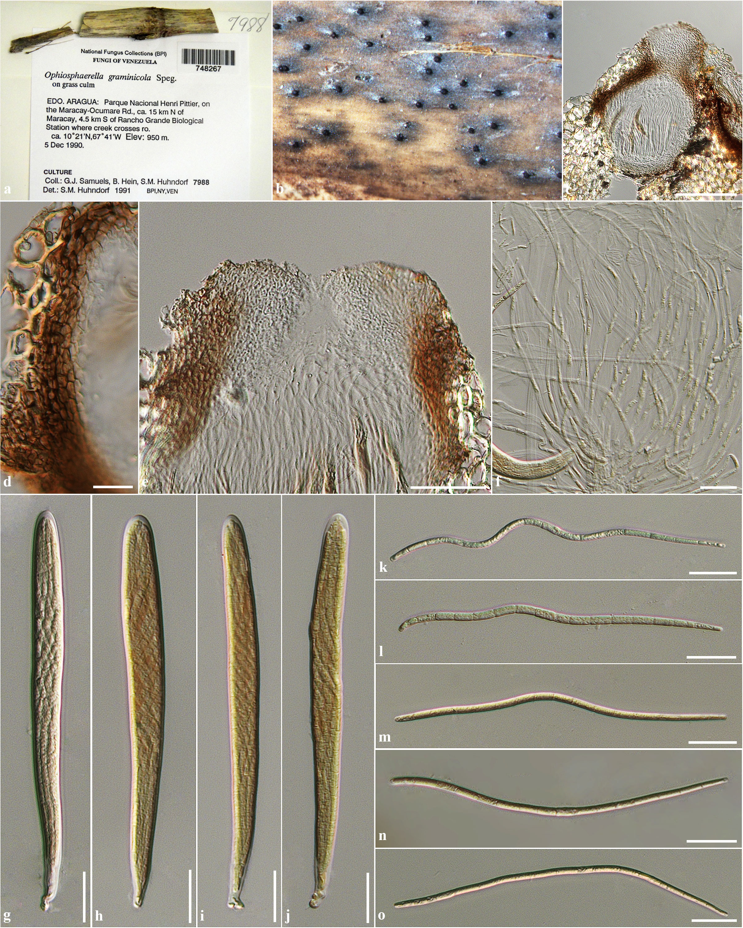
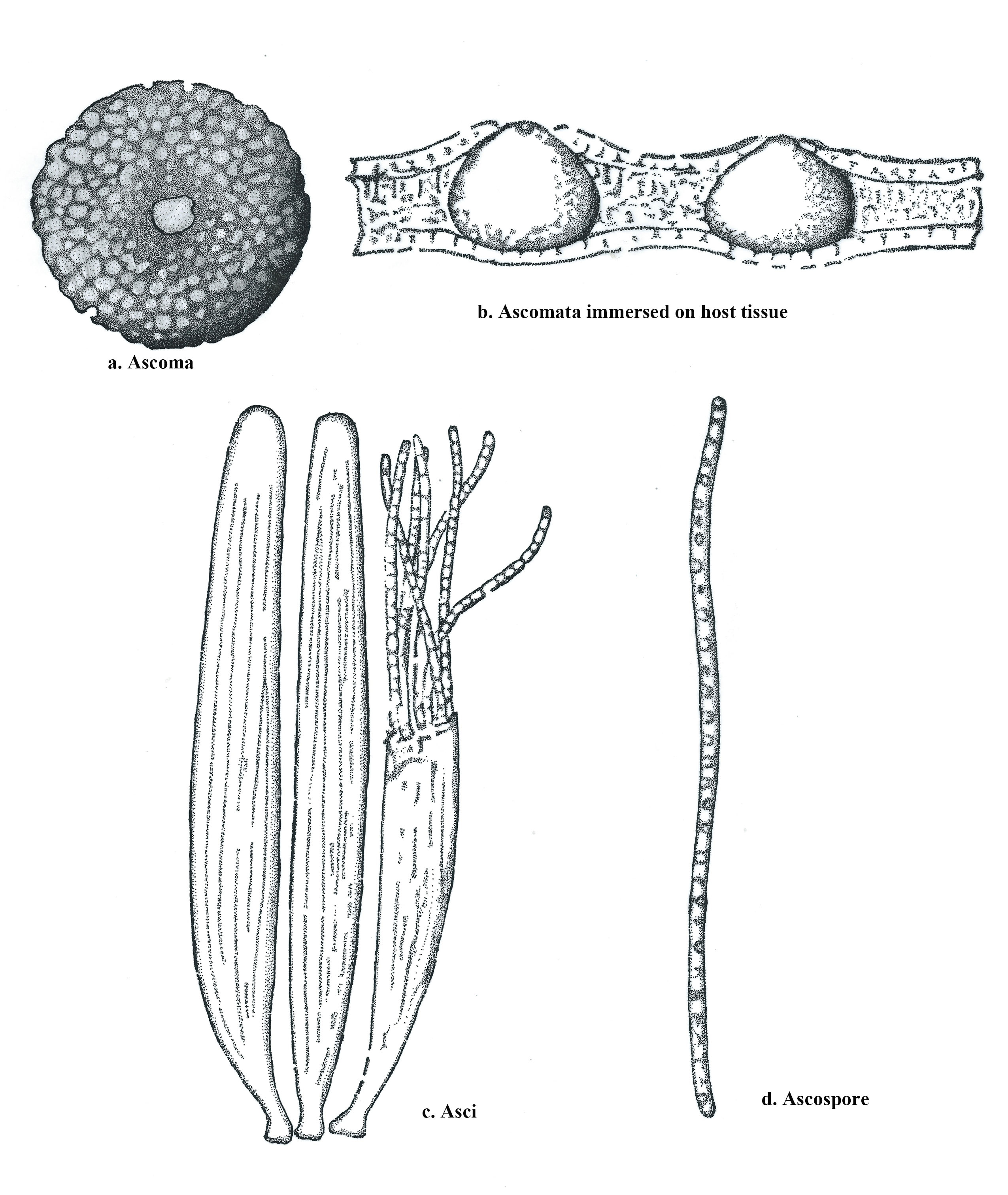
Fig. 17 Ophiosphaerella graminicola (iconotype) redrawn from Spegazzini (1909). a Ascoma. b Ascomata immersed on host tissue. c Asci. d Ascospore.