Fomitiporia atlantica Alves-Silva, Reck & Drechsler-Santos
Index Fungorum number: IF 551915 Facesoffungi number: FoF 01831
Etymology: referring to the vegetacional type where the fungus was found, the Atlantic forest.
Holotype: FLOR 58554.
Basidiomata perennial, pileate, sessile and mostly broadly attached, semicircular, solitary to imbricate, then with the different pilei fusing, with a nodulous aspect when emerging from the wood, obtriquetrous to obungulate, also triquetous, projecting 12.5–51 mm, 21–66 mm wide and 20–82 mm thick at the base, woody consistency when dried; pileus glabrous, concentrically zonated with multiple narrow bands, slightly sulcate, faintly cracked when old, dull, when fresh pilear surface greyish brown 11E3, violet brown11F4 to dark brown [7 F(6–8)], upon dried brown[6 E(5–8)]to olive brown [6 F(4–8)] when young [6 E(5–8)], becoming dark brown [6 F(5–8)]; margin finely velutinous, round, folded, thick, 3.5–19 mm thickness, sterile,olive brown [6 F(4–8)], yellowish brown to brown [5 DEF(6–8)]; pore surface light greyish brown (5D8) when young, greyish brown to cinnamon; pores rounded to angular, 6–8( –9) per mm, (60–) 70 –110 (–120) μm diam. (mean=89μm); dissepiments entire, (30–) 40 – 120μm (mean=67μm) thick; tubes distinct to mostly indistinctly stratified, with several layers (up to 15 layers in the oldest basidioma), those interleaved with context layers usually thicker (up to five times), individual tube layers relatively thin, sometimes difficult to distinguish, up to 2 mm tall, brown [5 EF (4–5)] to grayish brown (5E3), the older layers filled with whitish mycelium; context simple, up to 20 mm thick, concentrically zonate, sometimes constituted by extremely thin black lines (invisible to the unaided eye) that made the separation between growth layers of the context, with dense texture and woody consistency, golden to brownish yellow [5 BC (7–8)], with a distinct dark line at the surface, which is dark brown when young, becoming black, sometimes with a resinous aspect. Hyphal system dimitic in all parts; generative hyphae simple septate, hyaline to pale yellow, sparingly branched,2–3 μm diam; skeletal hyphae golden brown to reddish brown, unbranched, thick-walled, rarely with locals welling up to 8μm, in the context 4–5(–5.5) μm diam., the lumen 1.5–3 μm wide, in the hymenophoral trama 4–5(–6) μm diam., the lumen 1.5–3(–4) μm wide. Hymenium: hymenial setae absent, other sterile elements presents (as basidioles), hyaline, thin-walled; basidia subglobose to globose, hyaline, tetrasporic, 9–11×7–8 (mean=9.5×8μm) Q=1 –1.3μm (meanQ=1.18μm); basidiospores subglobose, globose to obovoid, with the wider portion displaced towards the apex, (4.5–)5 –5.5 (–6)×4–5.5μm (mean=5.1×4.8μm) Q=1 – 1.25μm (meanQ=1.08μm)(n=40), hyaline, strongto weakly dextrinoid, cyanophilous, thick-walled, smooth.
Material examined: BRAZIL, Santa Catarina, Blumenau, Parque Natural Municipal São Francisco de Assis, 26°55′17″S 49°04′18″W, on dead cut tree, 21 November 2014, G. AlvesSilva 640, (FLOR 58554, holotype); Ibid., on dead standing trunk, 15 September 2015, F. Bittencourt 507 (FURB 47591).
Notes: Fomitiporia atlantica is mainly characterized by the nodulose aspect of basidiomata with thick-rounded margin and darkness aspect of basidiomata when fresh, the narrowly zonated pilear surface, the zonation of the context (with variable presence of concentric thin black lines invisible to the unaided eye) and by the irregular layers of tubes; microscopically, the new species presents dimitic hyphal system and globose, subglobose to obovoid basidiospores with variable dextrinoid reaction. Fomitiporia atlantica shares with F. castilloi Decock & Amalfi the nodulous basidiomata (better observedinyoung specimens). However, F. castilloi is described by Amalfi and Decock (2013) from French Guiana as presenting distinct hymenial setae and slightly larger basidiospores in range and average (6.2×5.2μm), besides having a wider pilear zonation as well as an azonated context. Fomitiporia gabonensis Amalfi & Decock also presents imbricate basidiomata and variable dextrinoid basidiospores. Nevertheless, F. gabonensis was described by Amalfi et al. (2010) from Africa (Gabon) as presenting smaller basidiospores (4.7×4.1μm) and acute thinner margin. Besides the morphological evidences, F. atlantica is also supported by molecular results. The phylogenetic analysis (Fig. 128) showed the two specimens clustered together in a strong supported clade (BS=100, BPP=1). Fomitiporia atlantica forms a more inclusive clade with other two species, F. subtilissima (described below) and another undescribed species from Brazil (FLOR 58555). This clade displays nested vicinity to F. apiahyna sensu lato clade (Amalfi et al. 2014), appearing as a sister clade of this lineage. Fomitiporia atlantica differs from F. apiahyna (Speg.) Robledo, Decock & Rajchenb. sensu Amalfi and Decock (2013) mainly by its slightly smaller basidiospores (F. apiahyna=5.9×5.1μm) and pileus slightly sulcate and cracked, conspicuous features in F. apiahyna.
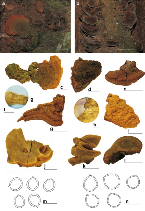
Fomitiporia subtilissima (FURB 47437) a Basidiomata in situ c Abmenial surface showing the concentric zonation and spathulate aspect of basidioma. Fomitiporia subtilissima (holotype) f, g Details of context and tubes f BlacklineatthesurfacegContext and tube layers j Hymenophoral surface m Basidiospores. Fomitiporia atlantica (FURB 47591) b Darkness aspect of basidiomata in situ e Abhymenial surfaceh Black line at the surface k Nodulous basidioma l Hymenophoral surface n Basidiospores. Fomitiporia atlantica (holotype) d Detail of slightly cracked abhymenial surface i Context and tube layers. Scale bars: a, b=50 mm, c –e, g and i, l=20 mm, f, h=2 mm, m, n=5μm