Tiarosporella dactylidis K.M. Thambugala, E. Camporesi & K.D. Hyde, sp. nov.
Index Fungorum Number: IF550768
Etymology: Referring to the host genus Dactylis.
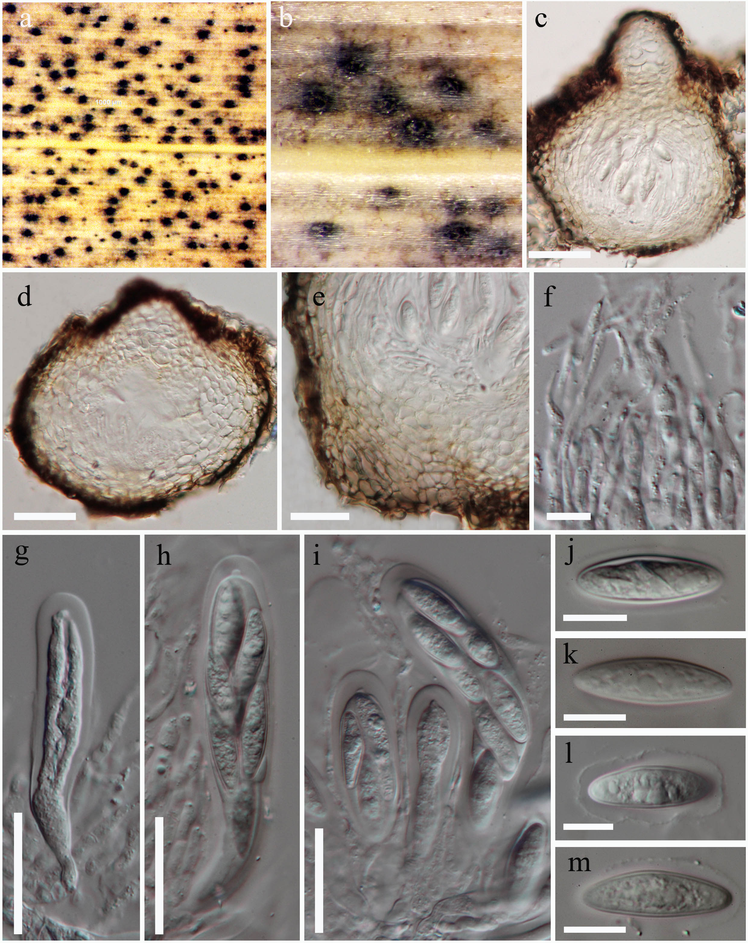
Saprobic on grasses in terrestrial habitats. Sexual morph: Ascomata 170–215 μm high × 160–250μm diam, pseudothecia, immersed to erumpent on host tissue, visible as black spots on host tissue, uniloculate, scattered or gregarious, globose to subglobose, ostiolate. Ostiole circular, central, papillate. Peridium up to 26–54 μm wide, broader at the base, comprising of 2 layers: outer layer of thin, small, brown to dark brown cells of textura angularis, inner layer of thick, large, hyaline to lightly pigmented, cells of textura angularis. Hamathecium comprising 2–3 μm wide, hyphae-like, hyaline, sparse pseudoparaphyses. Asci 80–160 × 17–21 μm (= 107 × 19.2 μm, n = 15), 8-spored, bitunicate, fissitunicate, clavate to cylindro-clavate, pedicellate, apically rounded with an ocular chamber. Ascospores 22.5–28 × 7–8.5 μm ( = 25.2 × 8 μm, n = 25), uni to bi-seriate in the upper half, uniseriate at the base, hyaline, aseptate, ellipsoidal to fusiform, usually wider in the middle, thick-walled, smooth-walled, surrounded by a mucilaginous sheath. Asexual morph: Conidiomata up to 500 μm diam, on pine needles, solitary or in groups, scattered, globose to sub globose, dark brown to black, immersed to semi-immersed on PDA, superficial on pine needles, pulvinate, unilocular, ostiolate. Conidiomatal wall several cell layers thick, with outer layers composed of dark-brown cells of textura angularis, becoming thin-walled and hyaline towards the inner region. Conidiophores up to 7 μm long, cylindrical, hyaline, unbranched. Conidiogenous cells 6.2–10 × 1.2–2.5 μm (= 7.8 × 1.8 μm, n = 15), formed from the cells lining the inner walls of the conidiomata, phialidic, fusiform to cylindrical, determinate, hyaline. Conidia 2–4 × 1–2.2 μm (= 2.7 × 1.4 μm, n = 40), solitary, ovoid, straight, oval to ellipsoidal, producing conidia at their tips, smooth, hyaline, aseptate.
Culture characteristics: Colonies on PDA, covering 90 mm diam. petri dishes after 4 d in the dark at 25°C; circular, initially white, after 1 week becoming greyish brown to black; reverse grey to dark grayish green, white in the margin; flattened, fluffy, fairly dense, aerial, smooth surface with crenate edge, filamentous and conidia produced on pine needles after 3 weeks at 18°C.
Material examined: ITALY. Teodorano – Meldola (province of Forlì-Cesena
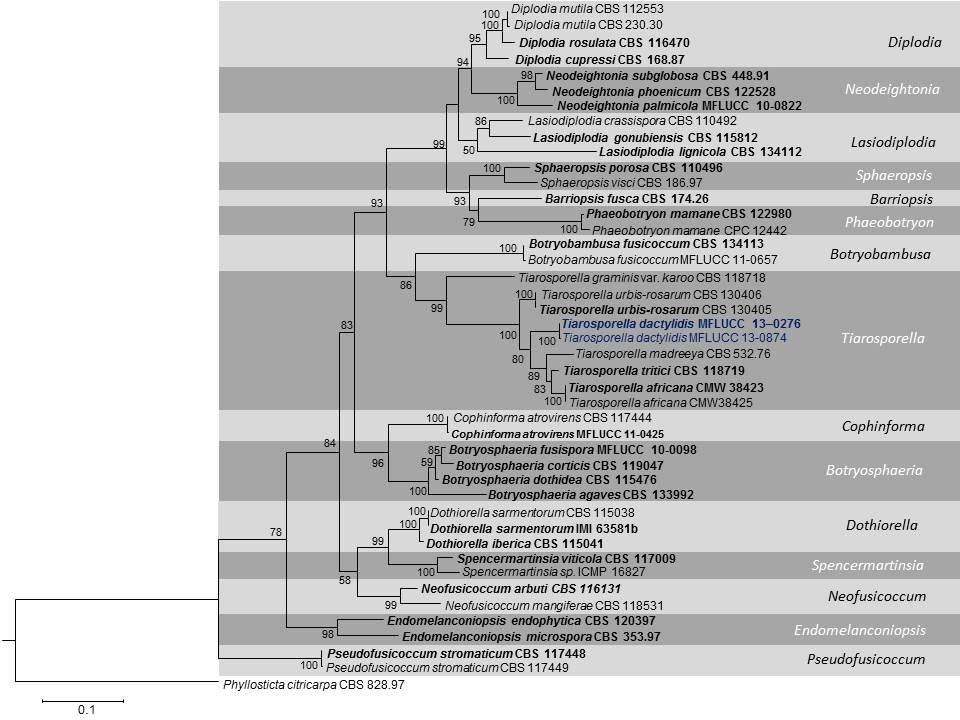
Notes: The sexual morph of Tiarosporella dactylidis is morphologically similar to Botryosphaeria in having globose ascomata with a central ostiole, a two-layered peridium, hyphae-like pseudoparaphyses and hyaline, aseptate, fusoid to ovoid ascospores with a mucilaginous sheath. The asexual morph of Tiarosporella dactylidis we observed in the culture looks similar to the spermatial states of Botryosphaeria (Phillips et al., 2013) and differs from the other asexual morphs of Tiarosporella (Crous et al., 2006; Jami et al., 2012, 2014). Our strains of T. dactylidis (MFLUCC 13–0276 and MFLUCC 13-0874) clustered in the Tiarosporella clade with 90% bootstrap support and this is the first sexual report for Tiarosporella. However, the generic type of Tiarosporella, T. paludosa has no molecular data and needs recollecting and sequencing to establish a better phylogeny.
Fig. 1. Best scoring RAxML tree based on a combined dataset of ITS, LSU and EF1-α. RAxML bootstrap support values (equal or greater than 50 %) are given at the nodes. The tree is rooted to Phyllosticta citricarpa (CBS 828.97). Ex-type strains are in bold.
Fig. 2. Tiarosporella dactylidis – sexual morph (MFLU 14–0580, holotype). a-b. Ascomata on host surface. c-d. Section through ascomata. e. Peridium. f. Pseudoparaphyses g. Immature ascus. h-i. Mature bitunicate asci j-m. Ascospores. Note the inconspicuous mucilaginous sheath. Scale bars: c-d = 50 µm, e = 25 µm, f = 10 µm, g-i = 30 µm, j-m = 10 µm.
Fig. 3. Tiarosporella dactylidis – asexual morph (MFLU 14–0580, holotype). a-b. Conidiomata on pine needles in culture. c. Squash mount of conidiomata. d-e. Conidia developing on conidiogenous cells. f. Conidia. Scale bars: c = 50 µm, d-e = 20 µm, f = 10 µm, j-m = 10 µm.


