Seimatosporium pseudoglandigenum Wijayaw. & E. Camporesi, in Wijayawardene, Goonasekara, Camporesi, Wang & An, Mycosphere 7(2): 207 (2016)
Index Fungorum number: IF552047, MycoBank number: MB552047, Facesoffungi number: FoF 02076
Etymology: Named as it morphologically resembles Seimatosporium glandigenum
Saprobic or endophytic on leaves of Quercus cerris L. Sexual morph: Undetermined. Asexual morph: Conidiomata 150−300 μm diam., 100−150 μm high, acervular, unilocular, subglobose to globose, superficial, gregarious, dark brown to black, apapillate ostiolate. Conidiomata wall multi-layered, outer wall thick, composed of brown cells of textura angularis, inner wall thin, hyaline, composed of hyaline cells of textura angularis. Paraphyses absent. Conidiophores 5−30 × 2−4 μm, long, cylindrical, branched, hyaline, smooth-walled. Conidiogenous cells holoblastic, simple, integrated, determinate, hyaline. Conidia 15−23 × 5−8 μm (x = 19.14 × 6.35 μm, n = 20), obovoid to fusiform, or cymbiform, obtuse apex, straight to slightly curved, with 3 transverse septa, septa dark brown, constricted at the septa or continuous, eguttulate, medium brown to golden brown, with hyaline to sub-hyaline basal and apical cell, smooth-walled.
Material examined − Italy, Forlì-Cesena [FC] Province, near San Paolo in Alpe – Santa Sofia, on decaying leaves of Quercus cerris L. (Fagaceae), E. Camporesi, 15 December 2013, IT 1577, MFLU 16−0837, holotype.
Notes − We made several attempts to isolate this taxon in different media, but were unsuccessful. Hence, we compare our new collection with related species by host association (Sutton 1980, Nag Raj 1993, Farr and Rossman 2016). Taxa reported from Quercus species are summarized in Table 2. Moreover, we compared our collection with other Seimatosporium species with 3 transverse septa (Sutton 1980, Nag Raj 1993), but our new collection is morphologically distinct.
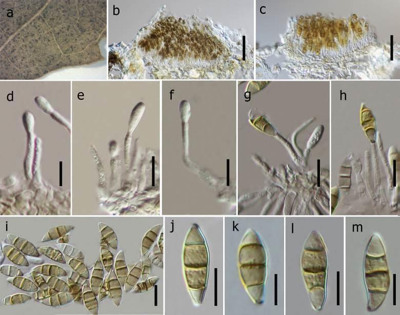
Fig 1 . Seimatosporium pseudoglandigenum (holotype, MFLU 16−0837). a Conidiomata on leaves of Quercus cerris. b, c Vertical sections of conidiomata. d-h Different stages of conidiogenesis. i-m Conidia. Scale bars: b, c = 100 μm, d-m = 10 μm.