Seimatosporium cornicola Wijayaw. & E. Camporesi, sp. nov,
Index Fungorum number: IF552048, MycoBank number: MB 552048, Facesoffungi number: FoF 02077
Etymology: Named after the host genus Cornus
Saprobic on dead branches of Cornus sanguinea. Sexual morph: Undetermined. Asexual morph: Conidiomata 330−400 μm diam., 220−250 μm high, acervular, superficial, solitary to gregarious, black, apapillate ostiolate. Conidiomata wall multi-layered, outer wall thick, composed of brown cells of textura angularis, inner wall thin, hyaline. Conidiophores 25−55 × 2−4 μm, long, cylindrical, branched, hyaline, smooth-walled. Conidiogenous cells holoblastic, simple, integrated, determinate, hyaline. Conidia 34−51 × 13−18 μm (x 41.86 × 16.1 μm, n 20), fusiform or obovoid, base truncate, straight, with 3 transverse septa, dark septa brown, constricted at septa, guttulate when immature, medium brown, with hyaline to subhyaline basal cell, smooth-walled, appendage absent.
Culture characteristics − On PDA slow growing, attaining a diam. of 2.5 cm in 7 days at 18 ºC, white to pale brown from above, greyish white from below, with sparse mycelium, flat, margin uneven.
Material examined − Italy, Forlì-Cesena [FC] Province, Camposonaldo – Santa Sofia, on dead branch of Cornus sanguinea L. (Cornaceae), E. Camporesi, 17 March 2012, IT 171 (MFLU 16−0701, holotype); ex-type living cultures MFLUCC 14−0448, GUCC IT 171.
Notes − Several Seimatosporium species have been recorded from Cornus spp. (Sutton 1980, Nag Raj 1993, Senanayake et al. 2015, Farr and Rossman 2016) (Table 3). In phylogenetic analyses, the new collection clusters with Seimatosporium pseudocornii (MFLUCC 13−0529) with high bootstrap values and PP values (86% and 1.00, respectively). However, in conidial morphology, both species are distinct (see Table 3). Seimatosporium pseudocornii has shorter conidiophores than in S. cornicola (5−30 μm vs. 25−55 μm). Hence, we introduce a new scientific name to accommodate our new collection.
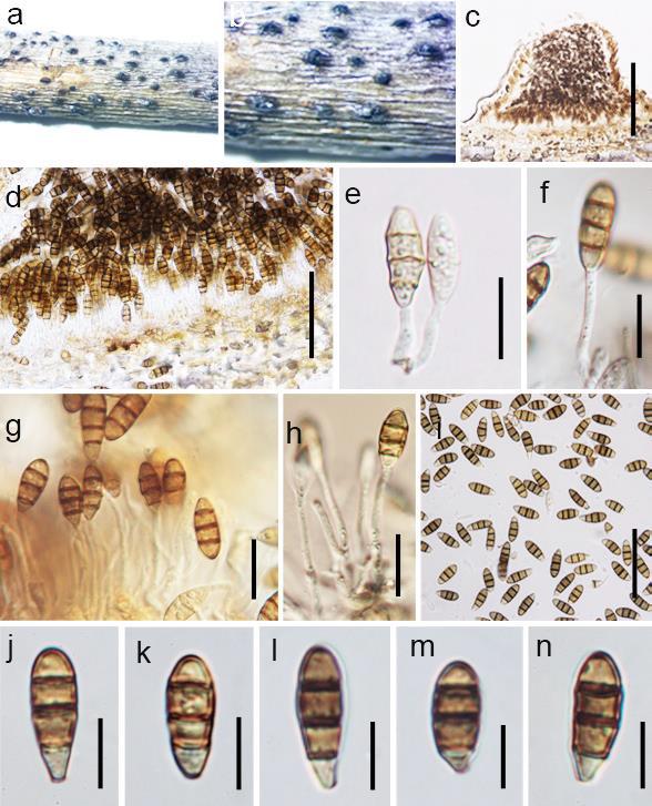
Fig. Seimatosporium cornicola (holotype, MFLU 16−0701). a, b Conidiomata on dead branch of Cornus sanguinea. c, d Vertical sections of conidiomata. e-h Developing conidia attach to conidiogenous. i-n Conidia. Scale bars: c = 200 μm, d = 150 μm, e-h = 30 μm, i = 100 μm, j-n = 35 μm.