Roussoellopsis macrospora (I. Hino & Katum.) I. Hino & Katum., J. Jap. Bot. 40: 87 (1965)—FIGS. 5, 6
≡ Didymosphaeria macrospora I. Hino & Katum., Bull. Faculty of Agriculture, Yamaguchi University 10: 1193 (1959)
Index Fungorum number: IF 338655; Facesoffungi number: FoF 000023
Pathogenic on living bamboo stems with other fungi. Sexual state: Ascostromata 0.3–0.4 mm high, 1.2–2.3 mm diam., raised, black, low convex or dome-shaped, solitary, uni- to multi-loculate. Locules 220–290 µm high, 540–720 µm diam., immersed under ascostromata, convex to elongate with a flattened base, centrally ostiolate. Peridium 7.5–20 µm wide, composed of two types of pseudoparenchymatous cells, forming textura angularis towards apex with textura prismatica at the base, covered by wedge-shaped as besides the stromatic region, unequal thickness, with slightly thin at the base of ascomata. Hamathecium comprising numerous, 1–2 µm wide, narrow, trabeculate, branched, anastomosing pseudoparaphyses, embedded in a gelatinous matrix. Asci (160–)180–200(–220) × (17‒)18–20(–25) µm (x̄ = 192.2 × 22.2 µm, n = 30), 8-spored, bitunicate, fissitunicate, clavate to cylindric-clavate, short pedicellate, apically rounded with distinct ocular chamber. Ascospores (28–)32–35.5(–37) × (10–)12–15 µm (x̄ = 32.2 × 12.4 µm, n = 30), uni-seriate or overlapping, rarely bi-seriate, broadly fusiform, brown to dark brown, 1-septate, constricted at the septum, rough-walled, echinulate, upper cell larger than lower cell, surrounded by distinct mucilaginous sheath. Asexual state: produced on sterilized bamboo pieces on WA. Conidiomata 250–380 µm high, 300–420 µm diam., visible as black dome-shaped on bamboo pieces, superficial, to embedded in agar, covered by vegetative hyphae, uni- to multi-loculate, solitary to gregarious. Conidiomata wall 30–70 µm wide, composed of dark brown cells of textura angularis, unequal thickness. Conidiophores (2–)6–20(–45) × (2–)3–5 µm, oblong to cylindrical, straight or curved, uni- to multi-septate, constricted at the septa, branching. Conidiogenous cells integrated, phialidic. Conidia (17–)20–25(–26) × (18–)19–24 µm (x̄ = 22.4 × 20.4 µm, n = 30), globose to subglobose, truncate at the base, hyaline when immature, with several small guttules, becoming dark brown with a large guttulate when mature, smooth-walled.
Culture characters:—Colonies on MEA 70–75 mm diam. after 4 weeks at 25–30◦C, white at the edge, yellowish white with small water drops in the centre; reverse white to pale yellowish at the edges, becoming dark greenish in the centre with slightly discontinued radiating outwards colony; dense, circular to irregular in shape, raised with flattened in the centre, smooth to slightly dull with entire edge, floccose to fluffy, slightly radiated in the upper part with strongly discontinue radiating in the lower part, non-pigmented.
Material examined: JAPAN. Tikugo Province: Dazaichu-tyô, Mt. Hôman, on dead culms of Phyllostachys bambusoides Sieb. & Zucc. (Poaceae), 9 September 1956, T. Hino (YAM, holotype); THAILAND. Chiang Rai Province: Muang District, Khun Korn Waterfall, on the living stem of bamboo (Poaceae), 21 June 2011, R. Phookamsak_RP0126 (MFLU 11-0244, epitype designated here iso epitype in PDD), ex-type living cultures = MFLUCC 12-0005 = ICMP.
Fig 1. Roussoellopsis macrospora. a. Ascostromata on the host surface. b. Section through dome-shaped ascostroma. c, d. Section through peridium. e. Asci embedded in pseudoparaphyses. f–h. Asci. i–m. Ascospores. n. Ascospore stained by Indian ink. o. Conidiomata forming on bamboo pieces on WA after 4 weeks. p. Section through conidioma. q. Conidiophores. r–s. Conidiogenous cells. t–x. Conidia. Scale bars: b = 200 μm, p = 100 μm, e–h = 50 μm, c, d, q, t = 20 μm, i–n, r, s, u–x = 10 μm.
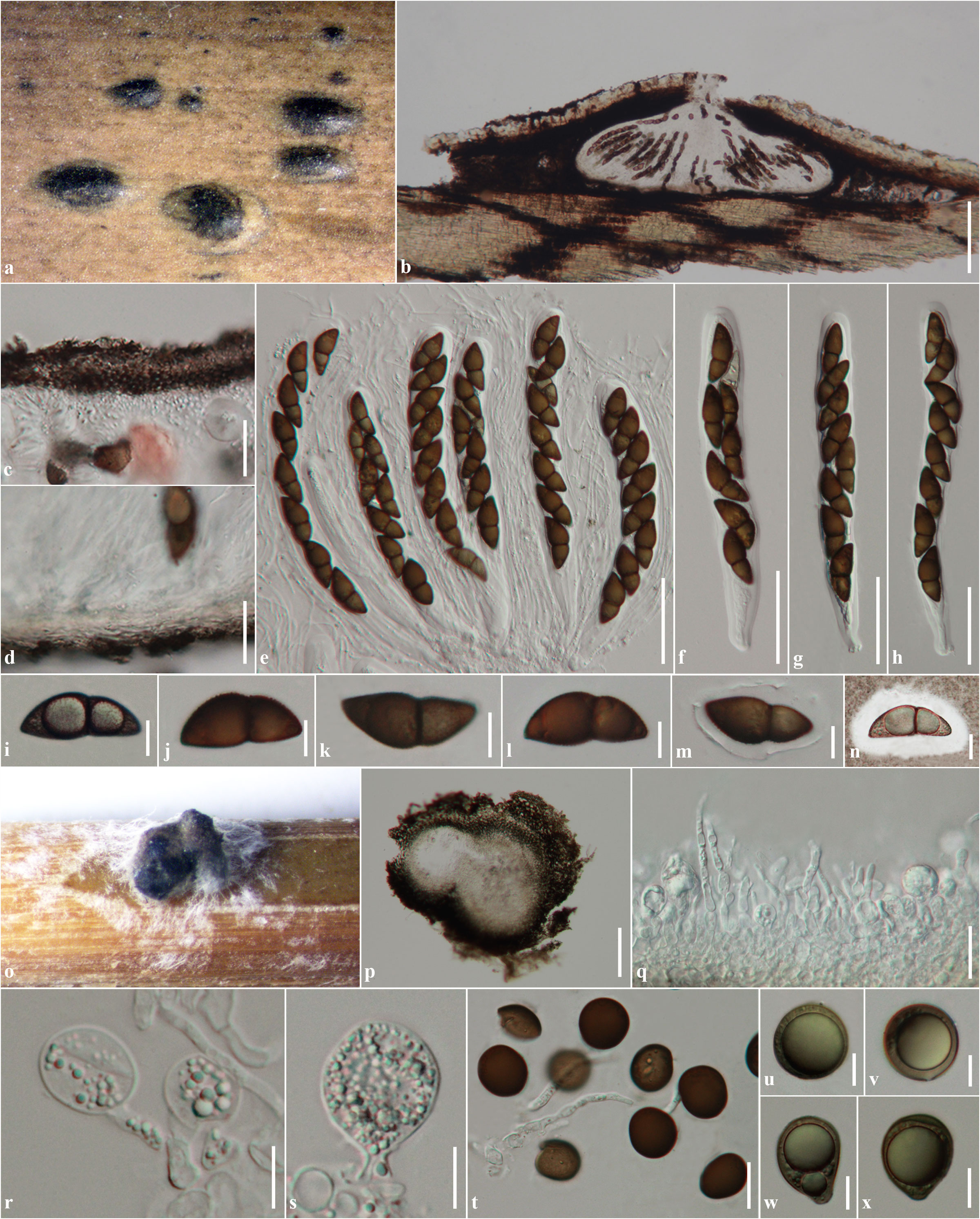
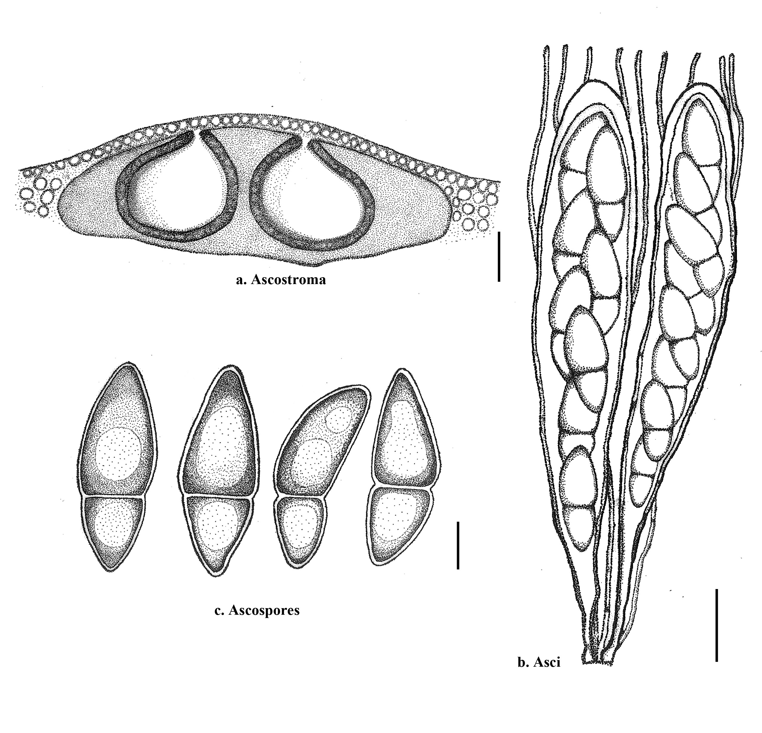
Fig 2. Roussoellopsis macrospora, re–drawn from Hino & Katumoto (1961). a. Section through ascostroma. b. Asci and pseudoparaphyses. c. Ascospores. Scale bars: a = 200 μm, b = 20 μm, c = 10 μm.