Paraleptosphaeria rubi Mapook, Camporesi, Ariyawansa & K.D. Hyde, sp. nov.
Index Fungorum number: IF551466, Facesoffungi number: FoF 01159
Etymology: The specific epithet rubi is based on the host genus from which the fungus was isolated.
Holotype: MFLU 15-1127
Fungicolous or saprobic on dead branch ofRubussp.Sexual morph: Ascomata 150–200 µm high × 200–250 µmdiam. (x̅ = 180 × 215 µm, n = 5), immersed or erumpent, solitary or scattered, globose to subglobose, brown to dark brown, smooth.Ostiolewith protruding papilla with periphyses.Peridium 10–20 µm wide, consisting of a single stratum, comprising brown to dark brown, scleroplectenchymatous cells oftextura angularis. Hamathecium comprising 1.5–2 µm wide, cylindrical to filiform, septate, branched, cellular pseudoparaphyses. Asci60–80 × 10–15 µm(x̅= 75 × 12 µm, n = 10), 8-spored, bitunicate, cylindric-clavate, slightly curved, with a short, bulbous pedicel, apically rounded, with an ocular chamber. Ascospores 20–30 × (5–)9–15 µm (x̅ = 25 × 10 µm, n = 20), 1–2-seriate, overlapping, hyaline, ellipsoidal, 1-septate, deeply constricted at the septum, upper cell widest and tapering toward rounded ends, straight or slightly curved, guttulate, surrounded by hyaline, mucilaginous sheath. Asexual morph: undetermined.
Material examined: ITALY, Province of Forlì-Cesena
Notes: Paraleptosphaeriarubi sits wellwith the generic concept of Paraleptosphaeria in havingimmersed, subglobose ascomata, thick-walled peridium comprising dark brown cells of textura angularis with cellular pseudoparaphyses, and cylindric-clavate asci bearing fusiform, transversally septate ascospores.
Paraleptosphaeria rubi is phylogenetically closely related to the type species of Paraleptosphaeria, P. nitschkei. Paraleptosphaeriarubi differs from P. nitschkei based on the position of the ascomata on the host (immersed versus superficial), ascospore septation (1-septate versus 1–3-septate) and habit (fungicolous versus saprobic). Furthermore, our new taxa has a mucilaginous sheath surrounding the ascospores, whereas the spores of P. nitschkei lack a sheath (Hyde et al. 2015).
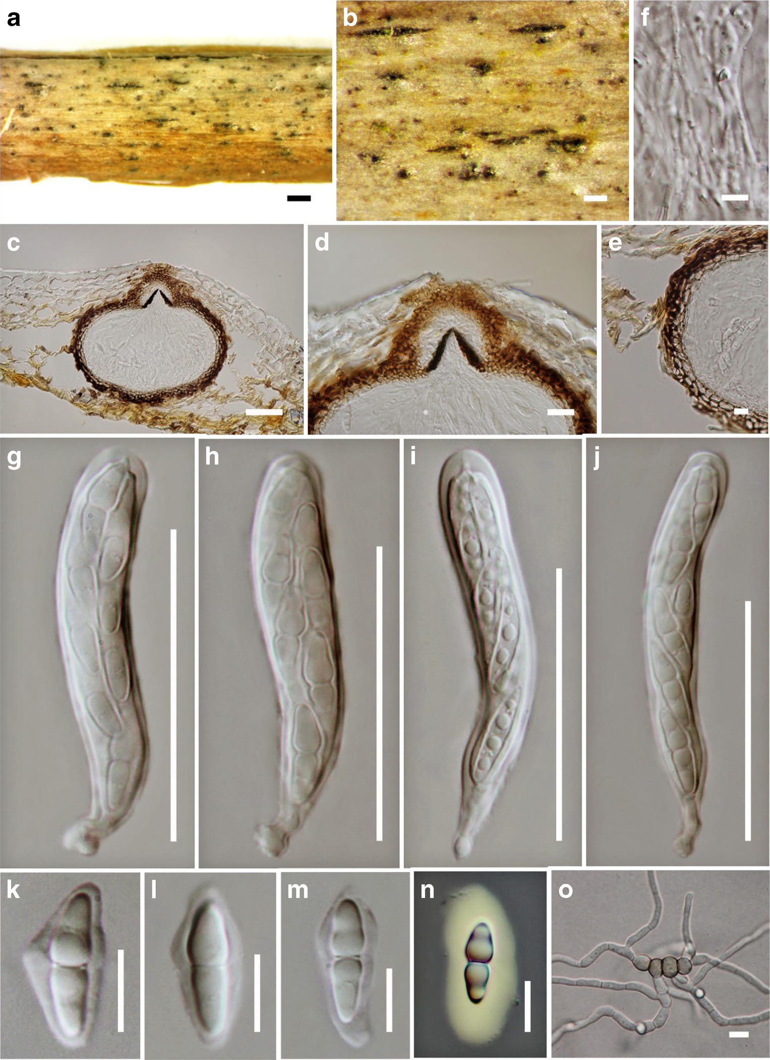
Fig.Paraleptosphaeriarubi a, b. Appearance of the ascomata on substrate. c. Section through ascoma. d. Ostiolar canal filled with periphyses. e. Peridium. f. Pseudoparaphyses. g–j. Asci k-m. Ascospores, N, surrounded by hyaline gelatinous sheath in Indian ink. o. Germinating ascospore. Scale bars: a = 500 µm, b = 200 µm, c, g–j = 50 µm, d = 20 µm, e, k–o = 10 µm, f = 5 µm.