Paraconiothyrium fuckelii (Sacc.) Verkley & de Gruyter, in Gruyter et al., Stud. Mycol. 75: 25 (2012) (Fig. 15).
Basionym: Coniothyrium fuckelii Sacc., Fungi venet. nov. vel. Crit., Sér. 5: 200 (1878).
Index Fungorum number: IF 564787; MycoBank number: MB 564787; Facesoffungi number: FoF 00055
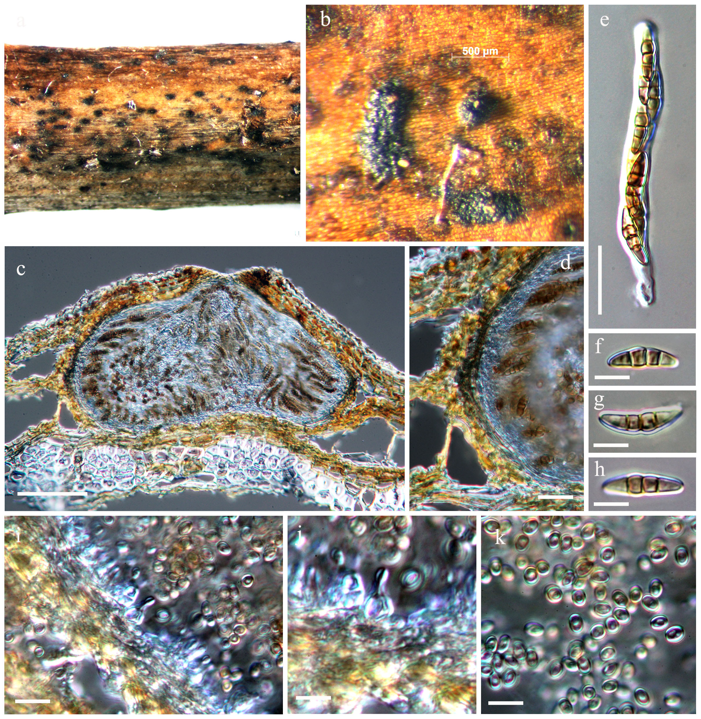
Saprobic on dead wood in terrestrial habitats. Sexual state: Ascomata 290–360 × 300–520 μm (x̄ = 300 × 430 µm, n = 10), solitary, scattered, or in small groups, immersed to erumpent, globose or subglobose, coriaceous, wall black, with or without papilla, ostiolate. Papilla small, with small ostioles. Peridium 15–40 μm wide, comprising one cell type of small, pigmented, thick-walled cells of textura prismatica to textura angularis. Hamathecium of dense, 1.5 μm broad, delicate pseudoparaphyses, 1-septate, branching and anastomosing between and above asci, embedded in mucilage. Asci 75–125 × 10–15 μm (x̄ = 90 × 12 μm, n = 10), 8-spored, bitunicate, fissitunicate, clavate, with a long, narrowed, furcate pedicel which is up to 45 μm long, and a low ocular chamber. Ascospores 15–18 × 5–7 μm (x̄ = 16 × 6 μm, n = 10), biseriate, narrowly ovoid to clavate, pale brown, 3-septate, constricted at the middle septum, smooth-walled. Asexual state: Conidiomata pycnidial 300–400 μm diam. and with a single cavity, more often eustromatic. Conidiomatal wall 3-layered, outer layer composed cells of textura angularis with somewhat thickened, brown walls, and an inner layer composed cells of textura angularis-globulosa with somewhat thickened, hyaline walls. Conidiogenous cells 4–10(–13) × 3–5 μm, discrete or integrated in short, simple, 1–2-septate conidiophores, broadly ampulliform to globose, holoblastic, often annellidic with 1 or 2 percurrent proliferations noticeable by the distinct scars on a somewhat elongated neck, hyaline. Conidia 3–4 × 2–3(–3.5) μm variable in shape, subglobose to ellipsoid or obovoid, rarely more cylindrical, initially hyaline, olivaceous-brown soon after secession, smooth, orange brown, aseptate.
Material examined – THAILAND, Chiang Rai, Bandu, on dead wood, 28 September 2012, K.M. Thambugala (MFLU 14-0305), living culture = MFLUCC 13-0043.
Fig. 1 Paraconiothyrium fuckelii (MFLU 14-0305). a–b Appearance of ascomata on the host surface. c Section of an ascoma. d Section of peridium. e Clavate ascus with a short, narrow pedicel. f–h Subglobose to ellipsoid or obovoid ascospores. i–j Conidiogenous cells. k Orange-brown conidia. Scale bars: c = 100 µm, d = 25 µm, e = 10 µm, f–k = 5 µm.