Ophiobolus disseminans Riess, Hedwigia 1(6): 27 (1854).
Saprobic on Cirsium (Asteraceae). Sexual state: Ascomata 140–390 μm high (excluding papilla), 160–340 μm diam., scattered or sometimes clustered, solitary to gregarious, semi-immersed, or erumpent through host tissue, visible as slightly raised, small, black dots on the host surface, uniloculate, subglobose to ovoid or ampulliform, glabrous, dark brown to black, ostiole central, with periphyses, short papillate. Necks 60–150 µm high, 85–140 µm diam, with thick-walled cells, arranged in a textura angularis, subglobose to oblong, dark brown to black, apically rounded or obtuse, coriaceous. Peridium 30–50 µm wide, thick-walled, of unequal thickness, slightly thick at the base, composed of several layers of broadly scleroplectenchymatous cells, outer layers consisting of several layers of brown to dark brown, thickened cells, arranged in a textura angularis, inner layers comprising several layers of hyaline, flattened cells, arranged in a textura epidermoidea to textura porrecta. Hamathecium composed of numerous, 2–3 µm wide, filamentous, broad cellular pseudoparaphyses, with distinct septa, embedded in mucilaginous matrix, anastomosing at the apex. Asci (156–)170–190(–203) × 10–12(–13) µm ( = 178.9 × 11.4 μm, n = 20), 8-spored, bitunicate, cylindrical to cylindric-clavate, short pedicellate, apically rounded with indistinct ocular chamber. Ascospores (125–)150–160(–163) × 2–3 µm ( = 152.8 × 2.9 μm, n = 15), fasciculate, in parallel or spiral, scolecosporous, filiform or filamentous, with two enlarged cells near the centre, but with 3–4 narrow cells in between the enlarged cells, brown, multi-septate, with up to 15 septa, not constricted at the septa, smooth-walled, separating into two part spores in the centre of the spores between the enlarged cells. Asexual state: Unknown.
Material examined: ITALY, Cassellas Vicinity, on dead stem of Cirsium arvense, 1853, Riess 1823 (BPI 629021, isotype); ibid on dead stem of Cirsium arvense, 1853, H. Riess, Hedwigia 1(6): Tab IV, (Fig. 8, iconotype) (1854).
Fig. 1 Ophiobolus disseminans (BPI 629021, isotype). a Herbarium label and specimen. b Ascomata on substrate. c Section through ascoma. d Section through peridium. e Papilla. f Asci with cellular pseudoparaphyses. g-i Ascus. j Ascospores in ascus. Note the swollen part of the ascospores (arrowed). Scale bars: c = 100 µm, d, e, f, g, h, i, j = 20 µm.
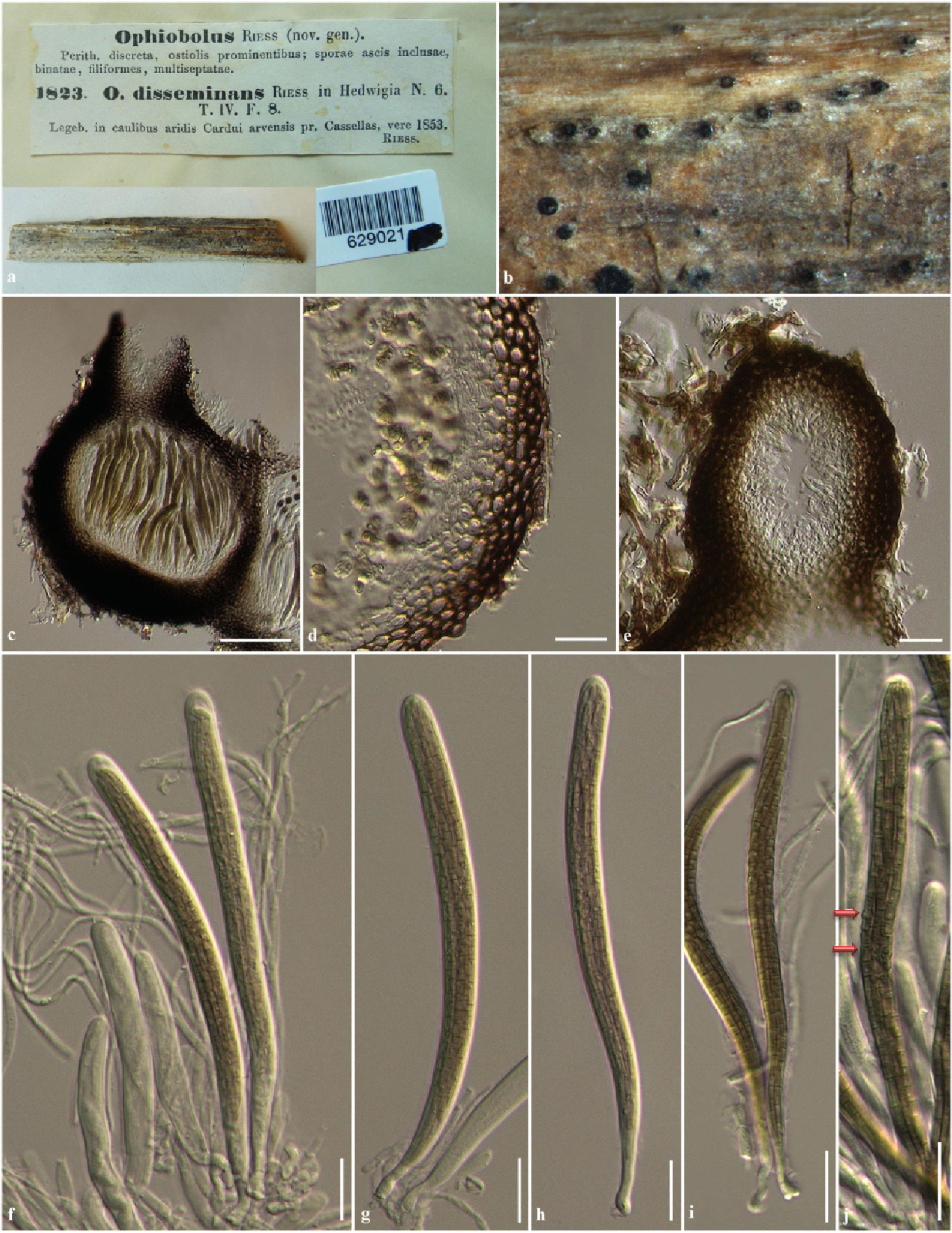
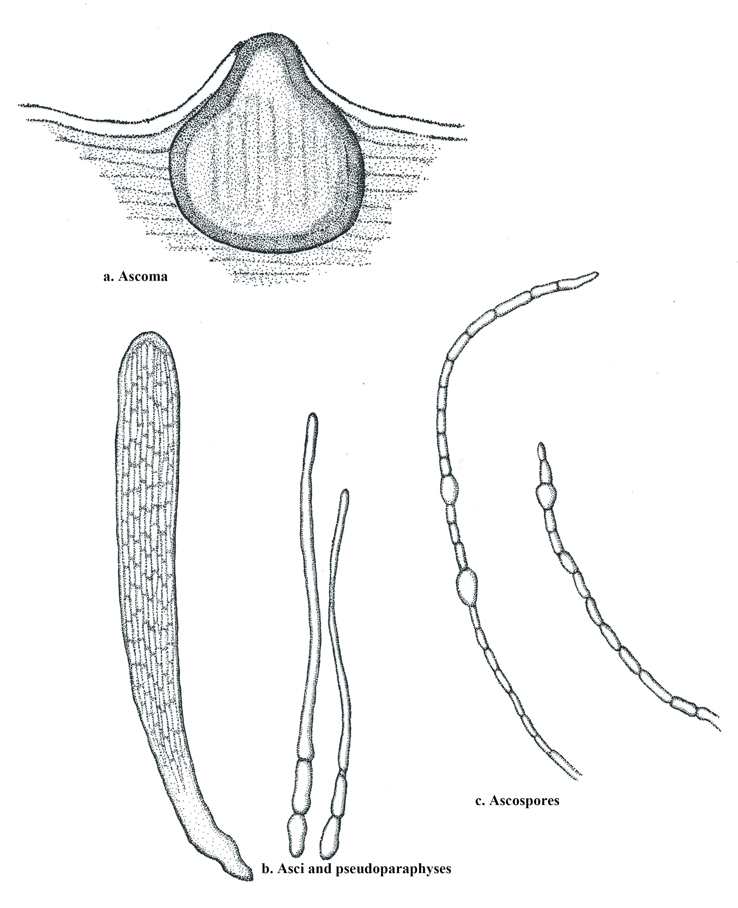
Fig. 2 Ophiobolus disseminans (iconotype) redrawn from Riess (1854). a Section through ascoma. b Ascus and pseudoparaphyses. c Ascospores.