Neokalmusia scabrispora (Teng) Kaz. Tanaka et al., comb. nov.,
Basionym:Leptosphaeria scabrispora Teng, Sinensia, Shanghai 4: 378 (1934).
Index Fungorum number: IF 550702; MycoBank number: MB 550702; Facesoffungi number: FoF 00052
Saprobic on culms of bamboo. Sexual state: Ascomata 200–300 × 130–500 μm, immersed under black clypeus-like structure composed of host epidermis and fungal mycelium, subglobose to oblong on host surface scattered to grouped. Ostiole absent or slightly papillate, about 85–100 μm long, with numerous periphyses. Peridium 7.5–20 μm thick at sides, composed of 3–6 layers of brown polygonal thin-walled cells of 5–10 × 2.5–6.5 μm, surrounded by wedge-shaped stromatic region (250–400 µm wide at sides) composed of vertically orientated hyaline cells of textura angularis. Hamathecium1–2 μm wide narrowly cellular, numerous, septate, branched and anastomosing, embedded in a mucilaginous matrix. Asci 123.5–160 × (15.5-)17–22 μm (= 142.1 ×1 8.8μm, n = 50), 8-spored, bitunicate, clavate, rounded at the apex and with an apical chamber, with a short stipe. Ascospores 29–40.5 × 7–10 μm (= 34.8 × 8.5 μm, n = 68), biseriate, fusiform to ellipsoid, slightly curved, 5 (rarely 7) -septate, with a median primary septum 0.48–0.53 μm (= 0.50 μm, n = 57) wide, slightly constricted at the septa, with third cell from the apex enlarged, penultimate cells shortest, brown to yellowish-brown, verrucose, with 10–20 μm wide sheath. Asexual state: unknown.
Material examined – JAPAN, Tochigi, Kanuma, near Ooashi river, on dead twigs of Phyllostachys bambusoides (Poaceae), 6 March 2003, N. Asama (KT 1023 = HHUF 28608), living culture CBS 120246 = JCM 12851 = MAFF 239517; Kagoshima, Kumagegun, Isl. Yakushima, Miyanoura river (riverbank), on dead twigs of Phyllostachys bambusoides (Poaceae), 17 Mar. 2007, K. Tanaka & H. Yonezawa (KT 2202 = HHUF 30013), living cultures = NBRC 106237.
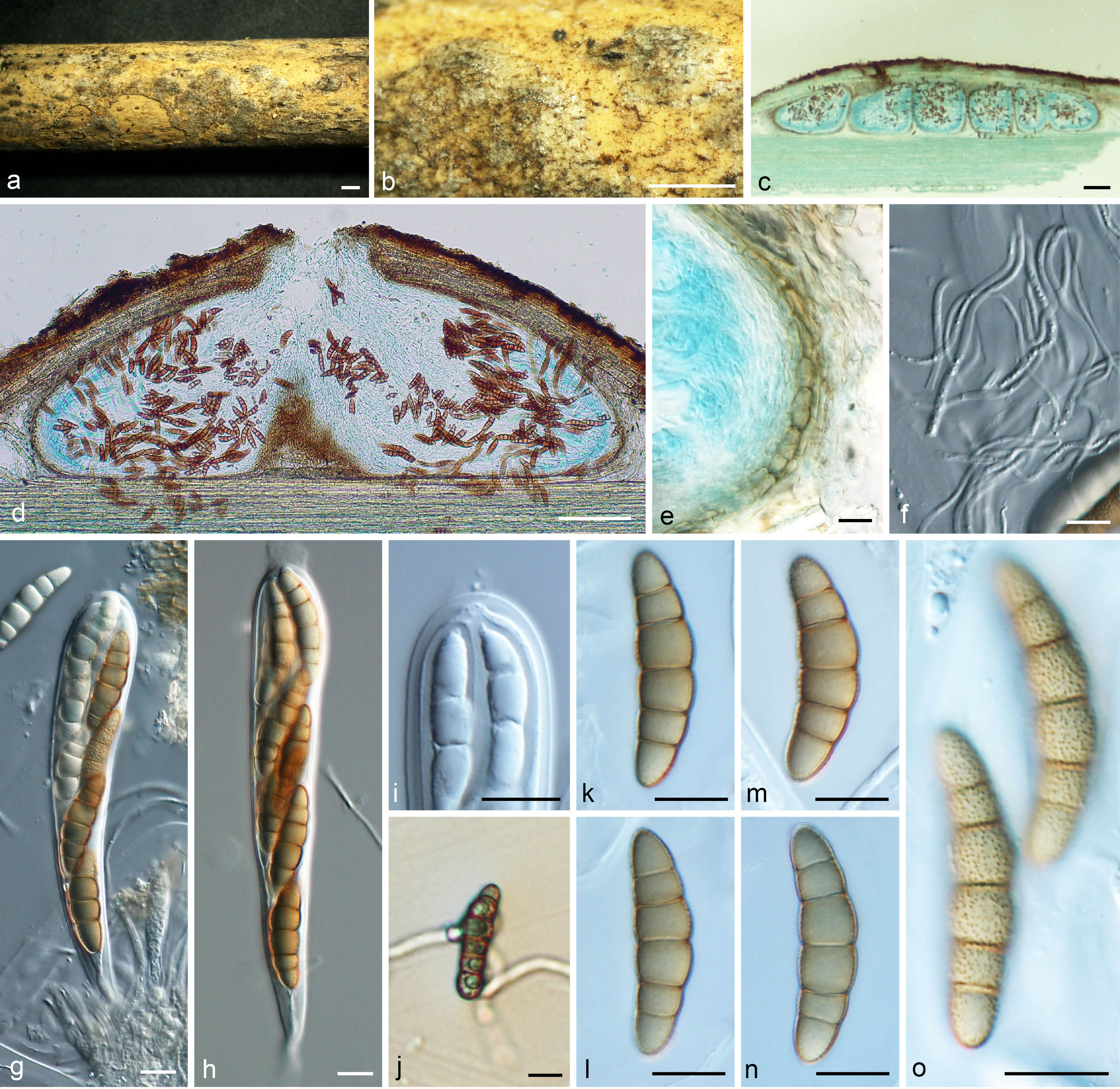
Fig. 1 Neokalmusia scabrispora (KT 1023). a–b Ascomata on host surface. c–d Vertical section through ascomata. e Section through peridium. f Pseudoparaphyses. g–h Asci. i Ocular chamber. j Germinating ascospore. k–o Ascospores. a–b, f–o from KT 2202, c–e from KT 1023. Scale bars: a–b = 1 mm, c–d = 100 µm, e–o = 10 µm.
Notes – This fungus was originally described as a species of Leptosphaeria (Teng 1934) and later transferred to Massariosphaeria (Shoemaker and Babcock 1989) or Kalmusia (Tanaka et al. 2005). Neokalmusia scabrispora, however, does not belong to the Leptosphaeriaceae typified by Leptosphaeria doliolum or to the Thyridariaceae encompassing the type species of Massariosphaeria (M. phaeospora) (Hyde et al. 2013). Neokalmusia scabrispora shares similar characters to Neokalmusia brevispora in having immersed ascomata under black clypeus-like, cellular pseudoparaphyses, 8-spored, bitunicate, clavate, asci rounded at the apex and fusiform to ellipsoid, brown to yellowish-brown, verrucose ascospores with a thick sheath. Neokalmusia scabrisporadiffers in the thickness of the peridium (7.5–20 μm versus 15–20 μm) and the number of septa in ascospores (5–7 versus 3).