Hymenochaete subporioides L.W. Zhou & Y.C. Dai
Index Fungorum number: IF551491 Facesoffungi number: FoF01005
Etymology: referring to the similarity to Hymenochaete porioides.
Holotype: BJFC011058 Basidiocarps annual, pileate, sometimes attached by a lateral tapering base, imbricate, corky and without odour or taste when fresh, hard corky and brittle when dry. Pilei dimidiate to usually fan-shaped, projecting up to 2.5 cm, 3 cm wide and 4 mm thick at base. Pileal surface cinnamon-buff, yellowish brown to brown, narrowly concentrically zoned in different shades, tomentose to velutinate; margin acute, convex when dry. Pore surface yellowish brown to dark brown; sterile margin distinct, yellowish brown, up to 1 mm wide; pores circular to angular, 6–8 per mm; dissepiments thin, entire. Context orange-brown, up to 3 mm thick, duplex, towards the tomentum separated by two black lines, lower and medium parts hard corky, upper tomentum soft corky. Tubes honey-yellow, paler than pores, up to 1 mm long. Hyphal system monomitic; generative hyphae with simple septa; tissue darkening and slightly swelling in KOH. Contextual hyphae in the lower dense context yellow, thick-walled with a wide lumen, unbranched, loosely interwoven, 3–5 μm in diam; hyphae in the two black lines dark brown, distinctly thick-walled with a narrow lumen, strongly agglutinated; hyphae between the two black lines yellow to brown, unbranched, parallel along the black lines, 3–4.5 μm in diam; hyphae in the upper tomentum brown, thick-walled with a wide lumen, unbranched, regularly arranged, 3–4.5 μm diam. Tramal hyphae varying from pale yellowish and slightly thick-walled to brown and thick-walled with a wide lumen, rarely branched, straight, parallel along the tubes, 2.5–4 μm diam. Setae frequent, distinctly subulate, arising from trama, most part embedded in hymenium, slightly curved at base, dark brown, thick-walled, 22–43×4–8 μm; cystidia and cystidioles absent; basidia more or less barrel-shaped, with four sterigmata and a simple septum at the base, 9–15×4–6 μm; basidioles in shape similar to basidia, distinctly smaller. Basidiospores oblongellipsoid to ellipsoid, hyaline, thin-walled, smooth, IKI–, CB–, (3.4–)3.7–4.3(−4.4)× (1.8–)1.9–2.4(−2.5) μm, L= 3.92 μm, W=2.18 μm, Q=1.8 (n=30/1).
Material examined: CHINA, Zhejiang Province, Yongjia County, Longwantan Forest Park, on angiosperm stump, 21 August 2011, Cui 10163 (BJFC011058, holotype), (IFP019138, isotype).
Notes: Hymenochaete subporioides resembles H. porioides by its pore size (H. porioides: 7–9 per mm, Ryvarden 2004; 6– 8 per mm, measured from LWZ 20140719-11) and duplex context separated by two black lines, but microscopically H. subporioides could be easily differentiated by its larger basidiospores (H. porioides: 2.5–3.5×1.5–2 μm, Ryvarden 2004; 2.5–3.1×1.5–1.9 μm, measured from LWZ 20140719-11). The ITS-based phylogeny also supports H. subporioides was separated from H. porioides.
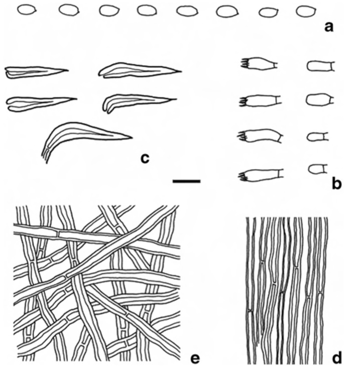
Hymenochaete subporioides microscopic structures (BJFC011058, holotype) a Basidiospores b Basidia and basidioles c Hymenial setae d Hyphae in trama e Hyphae in lower context Scale bars: a=5μm, b–e=10μm