Helicosporium vegetum Nees, Syst. Pilze (Würzburg): 68 (1816)
= Sphaeria cereaBerk. & M.A. Curtis, Grevillea4(no. 31): 108 (1876)
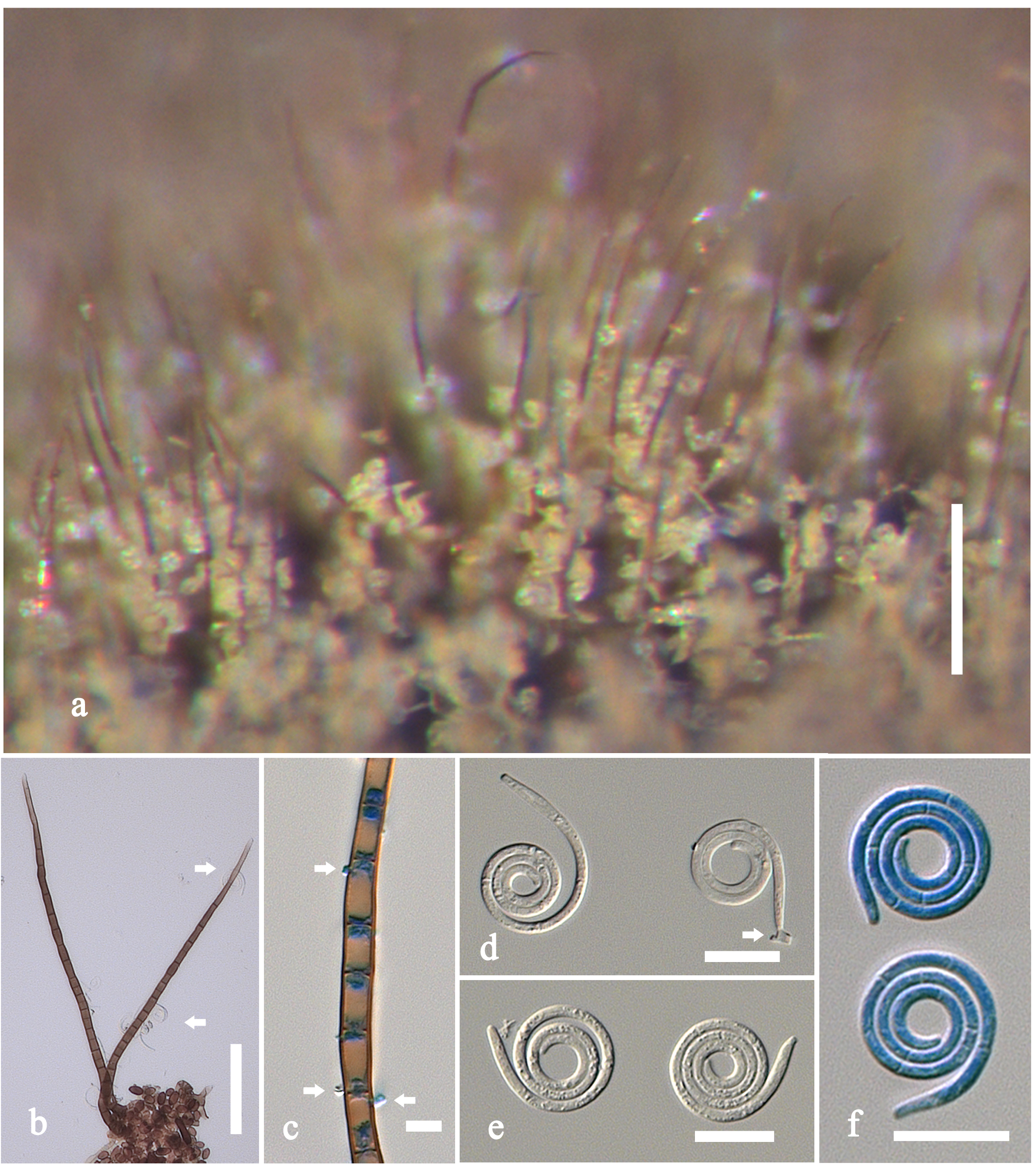
Saprobic on leaves, bark, and twigs, oron stromata of Diatrype stigma, Diatrypaceae (Xylariales), on decorticated or decaying wood, in terrestrial habitats, or submerged in freshwater, widespread in temperate to tropical regions. Sexual state: Ascomata180–211 µmhigh × 186–226 µm diam., superficial, solitary, scattered, globose-subglobose, bright yellow brown to yellow orange, collapsing when dry, darkened near ostiole. Peridium 23–26 µm wide, comprising several layers, composed of bright yellow cells of textura prismatica to angularis. Hamathecium comprising numerous, 1–1.5 µm wide, filiform, septate, branched, hyaline pseudoparaphyses. Asci 68–82(-92) ´ (7-)8–12 µm ( = 75 × 10 µm, n = 20), 8-spored, bitunicate, cylindric-clavate, apedicellate, thickened at apex, with an acute ocular chamber. Ascospores 35–46 ´ 3–5 µm ( = 39 × 4 µm, n = 10), biseriate, elongate-fusiform, tapering towards narrowly subacute ends, 8–9-septate, slightly curved, hyaline, smooth-walled. Asexual state: hyphomycetous, helicosporous. Conidiophores (98-)107–220 µm long × 3–5 µm( = 148 × 4 µm, n = 20),macronematous, mononematous, setiferous, erect, septate, unbranched, dark brown, fertile in middle, sterile,tapering toward narrow subacute at apex, smooth-walled,arising directly on substrate, from thick-walled, closely septate, repentent hyphae, crowded or in fascicles, glistening, light coloured. Conidiogenous cells polyblastic, intercalary, rarely terminal, with lateral minute denticles each with single conidium. Conidia 10–15 µm diam. and conidial filament 1–2 µm wide ( = 13 × 1.5 µm, n = 20), coiled 3½–4½ times, tightly to loosely coiled, rounded at apical end, truncate at base, 7–13-septate, slightly constricted at septa, hyaline, smooth-walled.
Notes: Von Höhnel (1919) transferred Sphaeria cerea Berk. & M.A. Curtis to Tubeufiaas T. cerea, based on bitunicate asci, and narrow elongate, multi-septate ascospores. Tubeufia cerea is often found on stromata of species belonging in Diatrypaceae (Xylariales). Morphologically, Tubeufia cereawas distinguished from all other species in the genus by its yellow brown to yellow orange ascomata (Bigelow and Barr 1963; Booth 1964; Samuels et al. 1979; Barr 1980). Booth (1964) provided a description and illustration of Tubeufia cerea based on the type specimen and provided a list of synonyms. The asexual state found near the ascomata were identified as Helicosporium vegetum Nees by Booth (1964), Samuels et al. (1979) and Barr (1980). Phylogenetic analysis links Tubeufia cerea with Helicosporium vegetum with high support (Tsui and Berbee 2006; Tsui et al. 2006) and this is supported in our study (Clade D, Fig. 2). Tubeufia cerea (Berk. & M.A. Curtis) Höhn. is therefore treated as a synonym of Helicosporium vegetum.
Material examined: USA, Virginia, Falls Church, on rotten wood, C.L. Shear, May 1936; Detr: J.A. Stevenson (BPI 447464, material of asexual state); Virginia, Shenandoah National Park, Beams Gap, on stromata of Diatrype stigma(Diatrypaceae,Xylariales), on dead wood, A.Y.Rossman(1925), 19 June 1983;Detr: A.Y. Rossman (BPI 1107327, sexual state of Tubeufia cerea).
Fig. 1 Helicosporium cereum (BPI 1107327 as Tubeufia cerea). a Ascomata on fungal stromata. b Section of ascoma. c Peridium. d Pseudoparaphyses. e, f Asci. g-i Ascospores. Scale bars: a=200µm, b=100µm, c=50µm, d=5µm, e-i=20µm
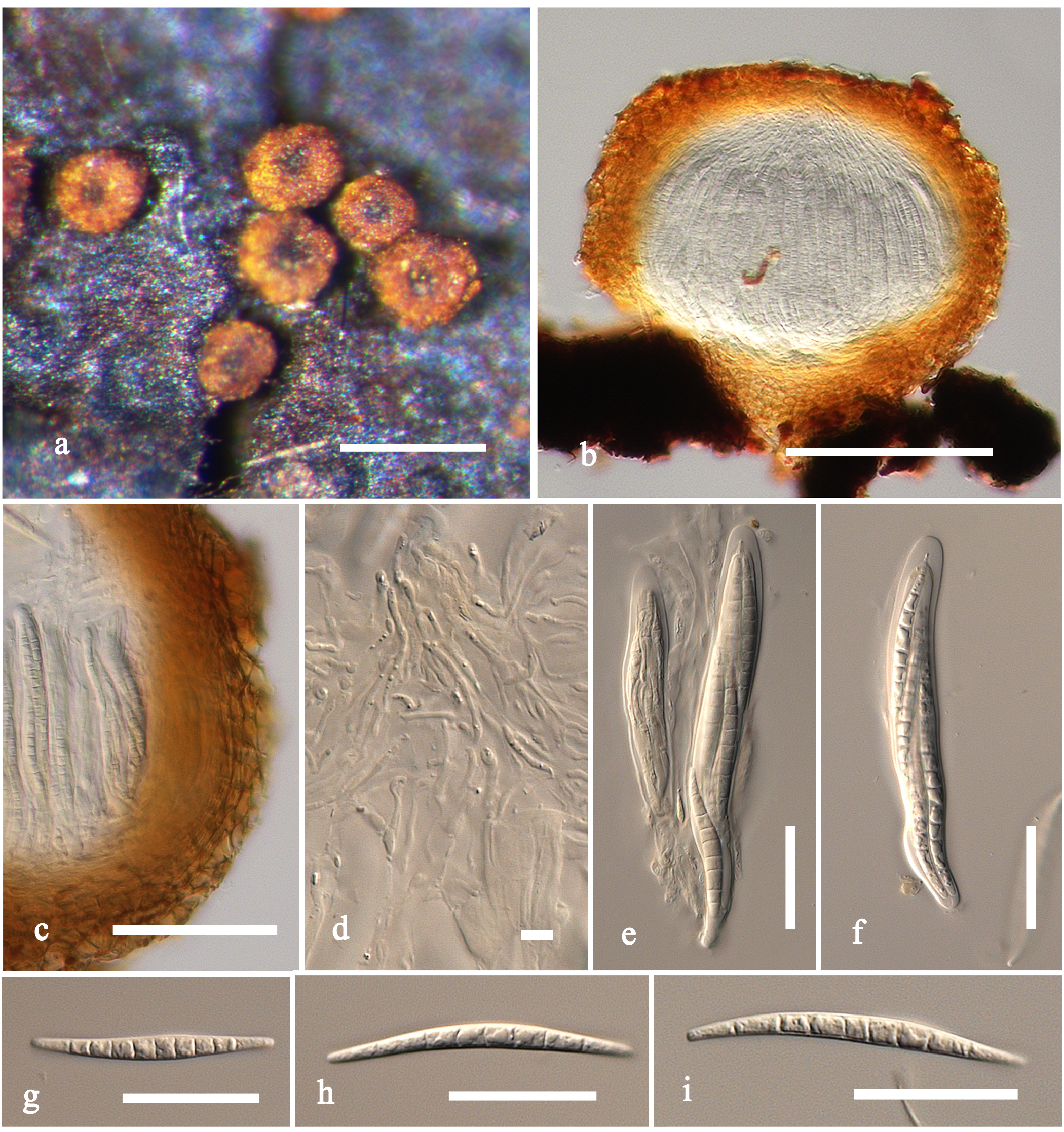
Fig. 2 Helicosporium vegetum (BPI 447464)a Conidiophores with conidia on natural substrate. b, c Conidiophores with minute denticles (arrows). d-f Conidia stained in cotton blue in Fig. f. Scale bars: a=100µm, b=50µm, c=5µm, d-f=10µm