Diaporthe collariana R. H. Perera & K. D. Hyde sp. nov., Indexfungorum number: IF554061
Etymology: Named after its prominently flared collarettes.
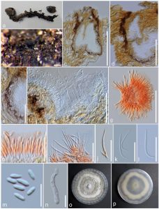
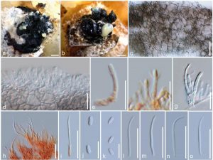
Saprobic on Magnolia champaca. Asexual morph from the natural substrate: Conidiomata 190– 325 μm wide, 310–550 μm high, pycnidial, eustromatic, subepidermal, semi immersed, scattered, globose to ampullifom or irregular, black, outer surface smooth, convoluted to unilocular, singly ostiolate, with prominent necks 150–290 μm long. Peridium 18–25 μm thick, 5–9 cells thick, consisting brown to hyaline cells of textura angularis. Conidial mass globose or sometimes exuding in cirrhi, white to pale-yellow. Alpha conidiophores 12.1–20.6 × 2.4–3.2 μm ( = 16.6 × 2.8 μm), densely aggregated, ampulliform to subcylindrical, rarely septate and branched, hyaline. Alpha conidiogenous cells 10–17 × 1.3–2.4 μm (x̅ = 13.7 × 1.8 μm) subcylindrical, tapering towards the apex, hyaline, with visible periclinal thickening, collarette prominent, up to 6 μm long, 5.7 μm wide. Alpha conidia 4.2–6.2 × 1.5–2 μm ( = 5.2 × 1.7 μm), less common than beta conidia, oblong to ellipsoidal, apex bluntly rounded, base obtuse to subtruncate, aseptate, straight, guttulate, hyaline, smooth-walled. Beta conidiophores 10.3–19 × 1.4–3.5 μm (x̅ = 14.6 × 2.6 μm), densely aggregated, subcylindrical, filiform or obconical, branched and septate, hyaline. Beta conidiogenous cells 3.8–14 × 1.4–2.2 μm (x̅ = 7.9 × 1.8 μm) subcylindrical, tapering towards the apex, hyaline, with visible periclinal thickening, collarette prominent, up to 6.6 μm long, 5.7 μm wide. Beta conidia 22–31.3 × 0.8–1.6 μm (x̅ = 27.7–1.2 μm), commonly found, straight, curved or hamate, hyaline, smooth-walled. Gamma conidia not observed. Asexual morph on PDA: Conidiomata 600–636 μm wide, 1045–1170 μm high, pycnidial, aggregated in small groups, globose to ampullifom, unilocular, black, with a prominent neck. Peridium consisting brown cells of textura angularis. Conidial mass globose or sometimes exuding in cirrhi, white to pale-yellow. Alpha conidiophores 12–20 × 2.4–3.2 μm (x̅ = 17.2 × 2.8 μm), densely aggregated, ampulliform to subcylindrical, rarely septate and branched, hyaline. Alpha conidiogenous cells 11.1–17 × 1.3– 2.4 μm (x̅ = 14.4 × 1.8 μm) subcylindrical, tapering towards the apex, hyaline, with visible periclinal thickening, collarette prominent, up to 3.5 μm long, 3.2 μm wide. Alpha conidia 4.7–5.6 × 1.7–2.2 μm (x̅ = 5.2 × 1.9 μm), less common than beta conidia, oblong to ellipsoidal, apex bluntly rounded, base obtuse to subtruncate, aseptate, straight, bi-guttulate, hyaline, smooth-walled. Beta conidiophores 13.2–20.8 × 1.3–4.1 μm (x̅ = 17.4 × 3.6 μm), densely aggregated, subcylindrical, filiform or obconical, branched and septate, hyaline. Beta conidiogenous cells 8.8– 13.4 × 1.7–2.3 μm (x̅ = 10.8 × 2.1 μm) subcylindrical, tapering towards the apex, hyaline, with visible periclinal thickening, collarette prominent, up to 3.5 μm long, 3.2 μm wide. Beta conidia 22–31.7 × 1.1–1.6 μm (x̅ = 28.8–1.3 μm), commonly found, straight, curved or hamate, hyaline, smooth-walled. Gamma conidia not observed. Sexual morph: Undetermined.
Culture characters: Conidia germinating on WA (Water Agar) within 12 h and germ tubes produced from one end. Colonies growing on PDA, reaching 6 cm in 7 days at 25°C, flat, initially white, aerial mycelium forming concentric rings with cottony texture, white to olivaceous, reverse zonate with white and ash-brown rings. Sporulate on PDA after 2 months incubation period in dark, at 25°C.
Material examined: THAILAND, Chiang Rai, Mae Fah Luang University premises, on dried fruits and pedicels of Magnolia champaca (L.) Baill. ex Pierre (Magnoliaceae), 17 August 2017, S. Boonmee, Fruit 3 (MFLU 17-2770, holotype), MFLU 17-2845 dried culture on PDA, ex-type living culture, MFLUCC 17-2636. (GenBank: LSU: MG806114)
Notes: Our new fungus Diaporthe collariana nested in between two D. subclavata strains and was more related to strain ZJUD83, which was collected from a fruit of Citrus maxima cv. Shatianyou in China, with very good support (Fig. 1). Nucleotide comparison reveals 5 (1.3%) differences in the ITS region, 10 (2.1%) in the TEF1 region, 11 (1.4%) in the TUB region. The extype strain of D. subclavata (ZJUD95) is the next phylogenetically closest isolate to Diaporthe collariana. Nucleotide comparison reveals 15 (3.8%) were distinct in the ITS region, 46 (9.7%) in the TEF1 region, 10 (1.2%) in the TUB region. CAL region is not available for D. subclavata strains in GenBank (Huang et al. 2015). Diaporthe collariana differs from D. subclavata in the presence of beta conidia. Furthermore, D. collariana produces prominent collarettes while collarets are absent in D. subclavata. On PDA, D. collariana produces smaller alpha conidia, which are oblong to ellipsoidal (4.7–5.6 × 1.7–2.2 μm), while D. subclavata produces fusiform to clavate conidia (5.5–7.2 × 2.2–2.9 μm) (Huang et al. 2015). The placement of D. collariana in between two isolates of D. subclavata is rather intriguing. However, by comparing available gene sequences of D. subclavata strains, we confirm that ZJUD83 is different from its extype ZJUD95. This is further discussed below.
Diaporthe micheliae, is another species which lacks molecular data in the GenBank, and was also isolated from the same host as D. collariana (Sankaran et al. 1987). However, D. collariana can be distinguished from D. micheliae by having prominent collarettes which are absent in D. micheliae, branched conidiophores (vs. simple conidiophores), and smaller alpha conidia (4.7–5.6 × 1.7–2.2 vs. 4.6–8.2(-11.5) × 2–2.8 µm) (Sankaran et al. 1987).
Fig. 1 Diaporthe collariana (MFLU 17-2770, holotype). a Herbarium material. b Conidioma on host substrate. c, d Section through conidiomata. e Peridium. f Alpha and beta conidia inside the same conidiomata. g, h Conidiophores with alpha conidia (in Congo red). i, j Conidiophores with beta conidia (in Congo red). k, l Beta conidia. m Alpha conidia. n Germinating conidium. o, p Colony on PDA. Scale bars: b, c = 200 µm, d = 50 µm, e–g = 50 µm, h–l = 20 µm, m, n = 10 µm.

Fig. 2 Diaporthe collariana (MFLU 17-2845) a, b Conidiomata on PDA. c Peridium in surface view. d Developing conidiophores with conidiogenous cells. e, f Conidiogenous cells with alpha conidia (in Congo red). g–i Conidiophores with beta conidia (h, i. in Congo red). j, k Alpha conidia. l–o Beta conidia. Scale bars: a, b = 1 mm, c = 20 µm, d–f = 10 µm, g = 10 µm, h, i = 20 µm, j, k = 10 µm, l–o = 20 µm.

