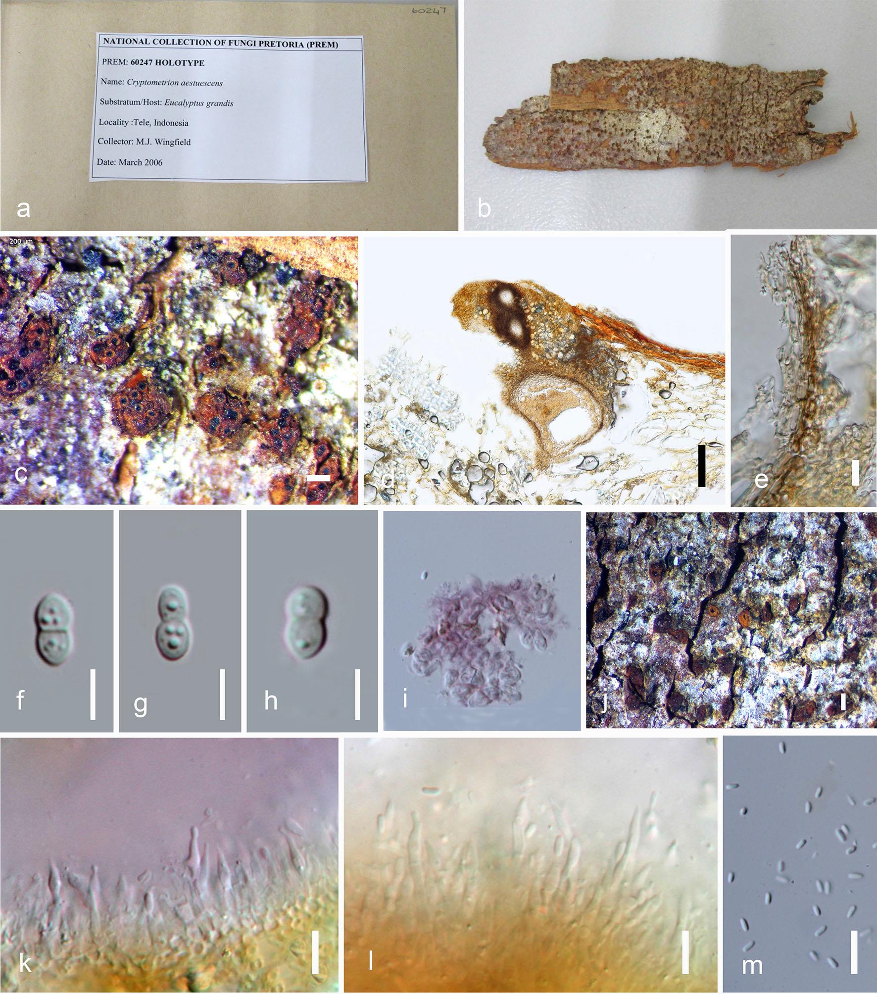
Cryptometrion aestuescens Gryzenh. & M.J. Wingf., Australas. Pl. Path. 39(2): 166 (2010).
Pathogenic on woody bark. Sexual morph: Ascostromata 60–130 mm high, 160–350 mm diam., on host gregarious or single, pulvinate to globose, semi-immersed in bark, orange, pseudoparenchymatous on edge of stroma, prosenchymatous in centre. Perithecia diatrypoid but can be forced by host tissue into a valsoid position, embedded in host tissue at base of stroma, fuscous black, necks emerge at stromatal surface as black ostioles covered with orange stromatal tissue to form papillae extending up to 230 mm above stromatal surface. Asci 25–32 × 5–7 μm , 8-spored, unitunicate, fusiform. Ascospores 6.5–7.5 × 2–3 μm , hyaline, fusoid to ellipsoid, one medial to submedian septum. Asexual morph: Conidiomata 80–180 μm high, 90–230 μm diam., part of ascostromata as conidial locules, or as separate structures, globose, semiimmersed, orange, uniloculate, with the same tissue morphology and stromatic structure as the ascostromata, spores expelled through opening. Conidiophores 8–13.5 lm long, occasionally with separating septa and branched, hyaline. Conidiogenous cells 1.5–2.5 μm wide, cylindrical or flaskshaped with apices attenuated or not, no paraphyses present. Conidia 3–5 × 1.5–2 μm , hyaline, cylindrical, aseptate, exuded as orange droplets (description based on Gryzenhout et al. 2010).
Material examined: SUMATRA, on bark of Eucalyptus grandis, PREM 60247 holotype.
Notes: Cryptometrion was introduced and typified by C. aestuescens. Cryptometrion is distinguished from other genera in Cryphonectriaceae based on its orange, limited stromatic tissue, uniseptate, fusoid to ellipsoid ascospores and the absence of paraphyses among the conidiogenous cells in the anamorph.
Fig. Cryptometrion aestuescens (PREM 60247). a Packet of herbarium. b Herbarium specimen. c Ascostromata on substrate. d Vertical cross section of ascomata. e Peridium. f–h Ascospores. i KOH reaction of stromatic tissues. j Conidiomata on substrate. k, l Conidiogenous cells and conidia. m Conidia. Scale bars: c, j = 200 μm, d = 100 μm, e–h, k–m = 10 μm.