Diaporthe acericola Dissanayake, Camporesi & K.D. Hyde, sp. nov., Index fungorum number: IF553186
Etymology: The specific epithet acericola is based on the host genus (Acer).
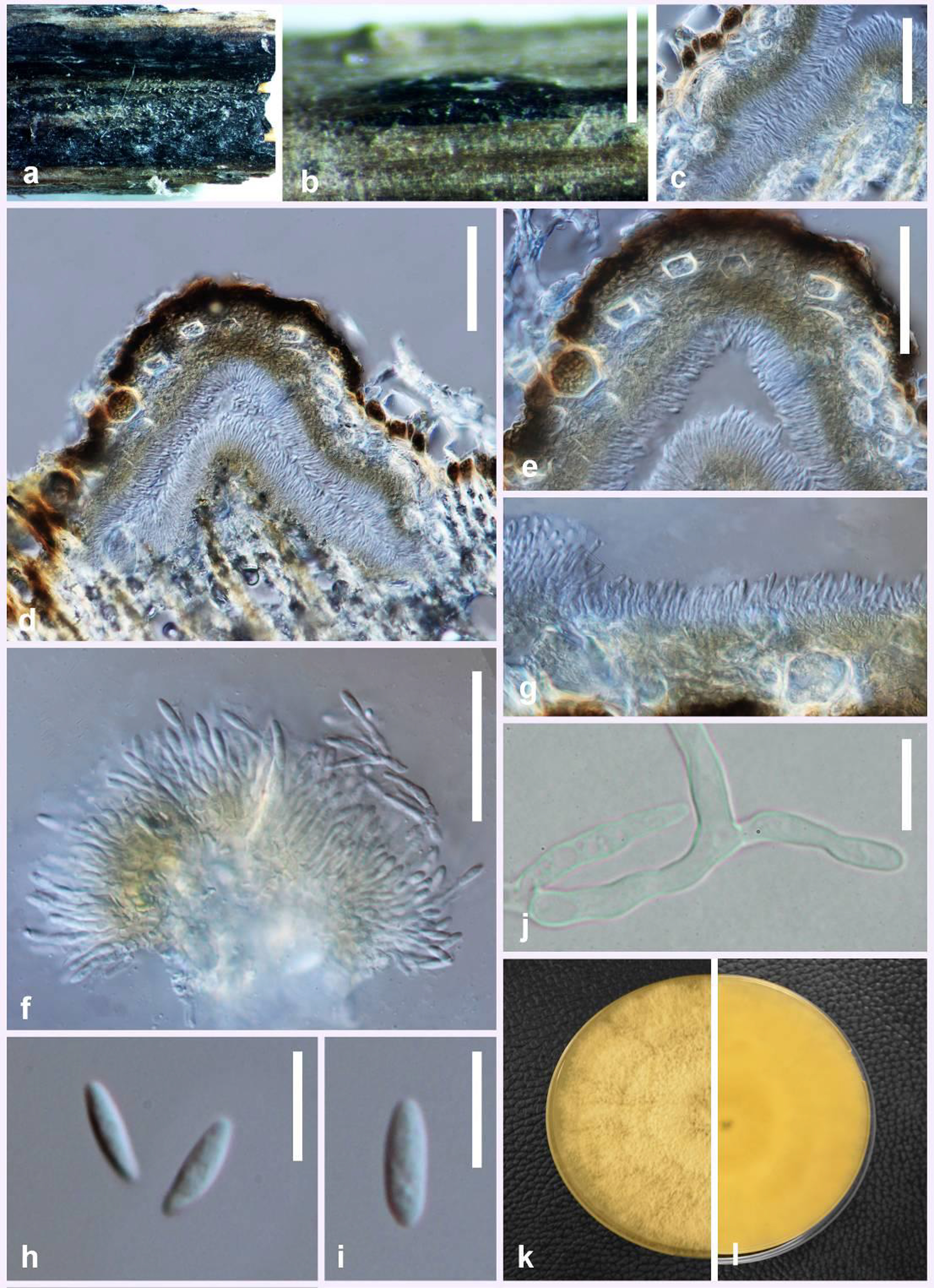
Saprobic on aerial branch and samaras of Acer negundo L. Sexual morph: Not observed. Asexual morph: Conidiomata up to 460 μm in diameter, 285 μm high, superficial, solitary, scattered on host, oval, black. Peridium 65–77 μm thick, inner layer composed of light brown textura angularis, outer layer composed of dark brown textura angularis. Conidiophores 21–35 ×1.5–2.5 μm (x̅ = 27 × 2 μm), cylindrical, aseptate, densely aggregated, straight or sinuous, terminal, slightly tapered towards the apex. Conidiogenous cells 10–15 × 2–3 μm, phialidic, cylindrical, terminal and lateral. Alpha conidia 9.7–13.5 ×3–4.5 μm (x̅ = 11 × 4 μm), hyaline, fusiform or oval, both ends obtuse. Beta conidia not observed.
Culture characteristics: Colonies on PDA covering entire Petri dishes after seven days at 25 °C, grey, with scant aerial mycelium; reverse fuscous black. Surface dirty white with profuse aerial mycelium, reverse umber.
Material examined: ITALY, Forlì-Cesena Province, San Colombano – Meldola, on dead aerial branches and samaras of Acer negundo (Sapindaceae), 22 January 2015, Erio Camporesi (MFLU 15-3254, holotype); ex-type living culture MFLUCC 17-0956.
Notes: Diaporthe acericola forms a sister clade to D. schoeni which is also a new species introduced in this study. However, the two species differed by 62 nucleotides in the concatenated alignment, of which 13 were distinct in the ITS region, 26 in the TEF region, 2 in the BT region and 21 in the CAL region. Morphologically, D. acericola differs from D. schoeni in having larger conidiomata and smaller conidia. Conidia of D. acericola are obtuse at both ends, while the conidia of D. schoeni are slightly acute and tapered at both ends.
Fig. Diaporthe acericola (MFLU 15-3254, holotype). a, b Conidiomata on host surface. d Cross section of conidioma. c,e Peridium. f, g Conidia attached to conidiogenous cells. h, i Alpha conidia. j Germinating spore. k, l Culture on PDA after one week. Scale bars: b = 0.5 mm, c–f = 100 μm, g–i = 10 μm.