Diaporthe schoeni Dissanayake, Camporesi & K.D. Hyde, sp. nov., Index fungorum number: IF553191
Etymology: The specific epithet schoeni is based on the host genus (Schoenus).
Saprobic on dead aerial stem of Schoenus nigricans L. Sexual morph: Not observed. Asexual morph: Conidiomata up to 210 μm in diameter, 110 μm high, immersed, solitary or gregarious, scattered on host surface, globose to oval, dark brown to black. Peridium 9–32 μm thick, inner layer composed of light brown textura angularis, outer layer composed of dark brown textura angularis. Conidiophores absent. Conidiogenous cells 21–35 × 1.5–2.5 μm (x̅ = 27 × 2 μm), cylindrical, aseptate, densely aggregated, straight or sinuous, terminal, slightly tapered towards the apex. Alpha conidia 11–14.5 × 2–3 μm (x̅ = 13.5 × 3 μm), hyaline, fusiform or oval, both ends slightly acute and tapered. Beta conidia 21–33 × 1–1.5 μm ( x̅ = 27 × 1.5 μm), rarely found among alpha conidia, hyaline, aseptate, filiform, hamate, tapering towards both ends.
Material examined: ITALY, Ravenna Province, Lido di Dante, on dead aerial stem of Schoenus nigricans (Cyperaceae), 1 May 2015, Erio Camporesi; (MFLU 15-1279, holotype).
Notes: We could not obtain a culture from single conidia. Therefore, fungal DNA was extracted directly from the conidiomata. Three isolates of D. schoeni were isolated from three different hosts, Carduus sp. (Asteraceae), Plantago sp. (Plantaginaceae) and Schoenus nigricans (Cyperaceae). However, any of those isolates were failed to germinate. Diaporthe schoeni occurs in a clade closer to D. acericola. Both species can be differentiated by smaller conidiomata and larger conidia of D. schoeni. Conidia of D. acericola are obtuse at both ends, while the conidia of D. schoeni are slightly acute and tapered at both ends. Phylogenetically, D. schoeni differs from D. acericola by 62 nucleotides in the concatenated alignment, of which 13 were distinct in the ITS region, 26 in the TEF region, 2 in the BT region and 21 in the CAL region.
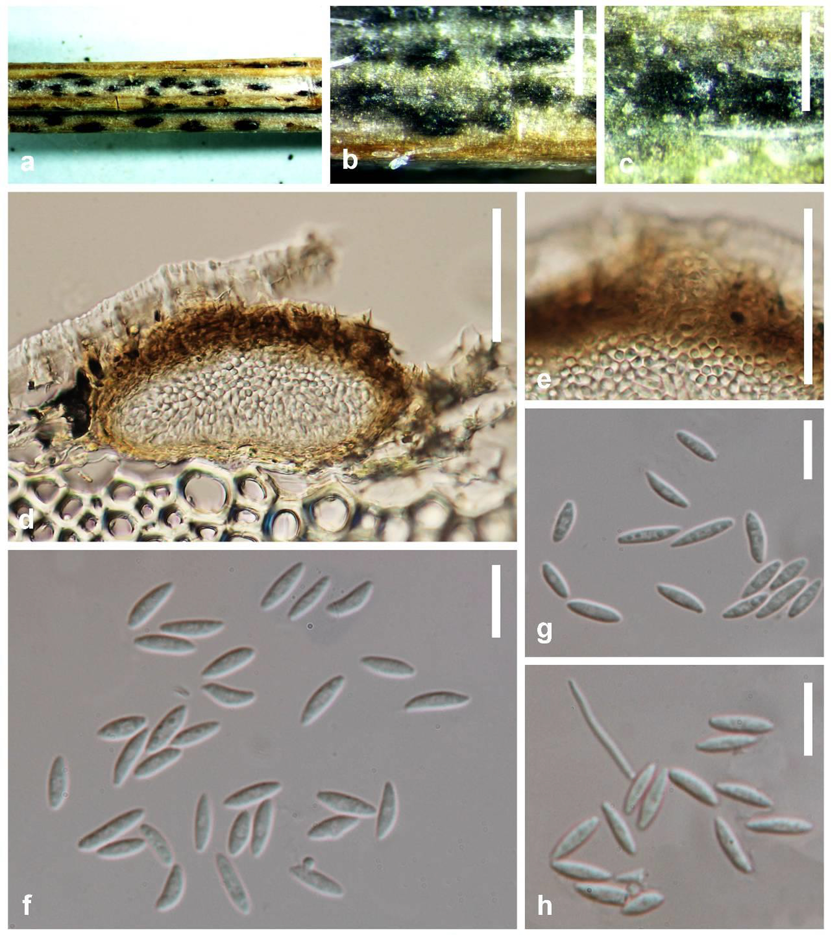
Fig. Diaporthe schoeni (MFLU 15-1279, holotype). a–c Conidiomata on host surface. d Cross section of conidiomata. e Peridium. f, g Alpha conidia. h Alpha conidia with a beta conidium. Scale bars: b, c = 1 mm, d,e = 100 μm, f–h = 15 μm.