Dactuliophora tarrii C.L. Leakey, Trans. Br. mycol. Soc. 47(3): 343 (1964).
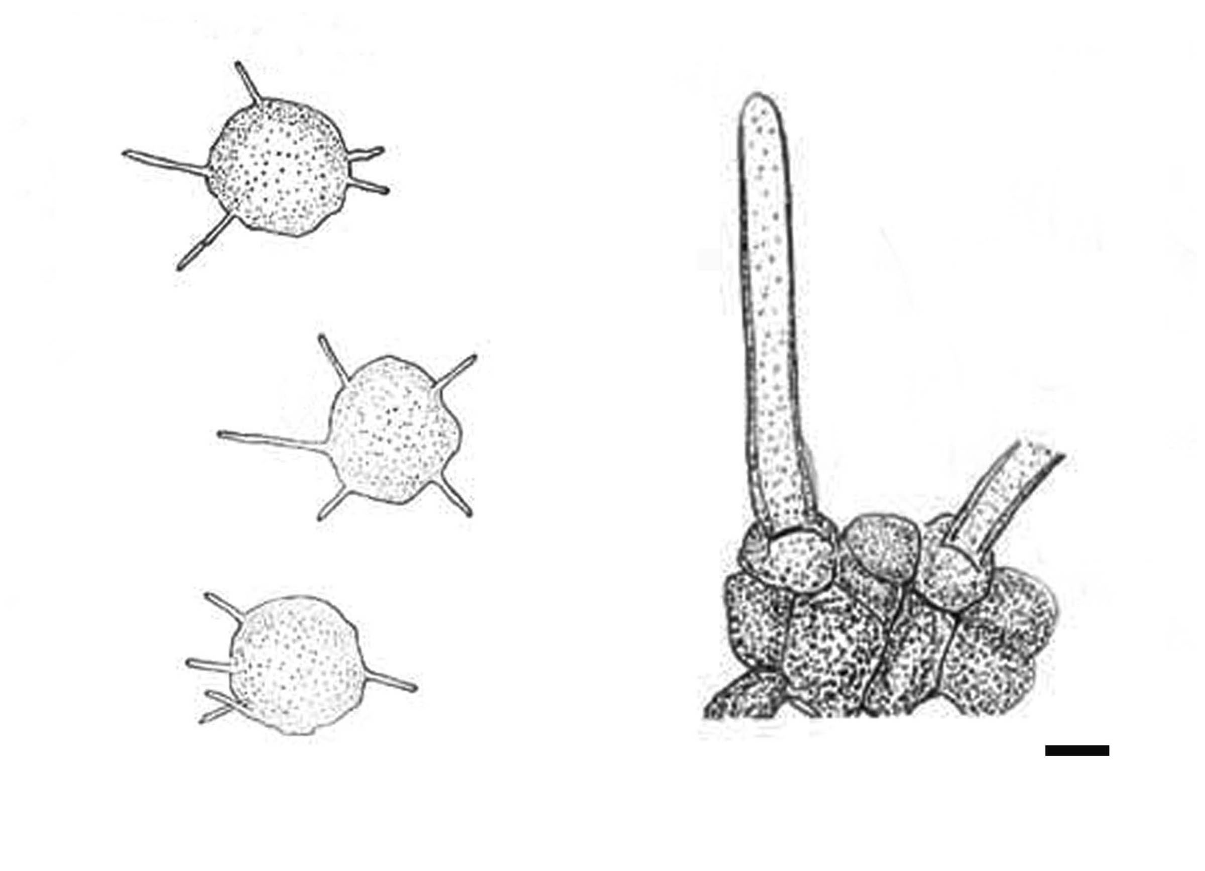
Parasitic on leaves. Leaf spots broadly zonate on upper leaf surface, rosette-like altering white and brown bands, lower surface densely covered with sclerotiophores and sclerotia, zonation less distinct. Mycelium immersed, hyaline, generally diffused throughout the tissues in the leaf spot and aggregated in plectenchymic masses beneath the sclerotiophores. Sclerotiophores consist of a fully erumpent ring of dematiaceous hyphae continuous with a subepidermal plectenchymic mycelial mass. Sclerotia 35–135 µm diam., scattered, specially on the paler parts of the leaf spot, generally hypophyllous but occasionally amphigenous, spherical to subspherical, bearing 0–10 (usually 3–7) scattered setae or sometimes glabrous, dematiaceous external cells (8–12 µm diam.), internal cells hyaline, undifferentiated. Sclerotial setae 3–4 µm diam., when present more or less cylindrical but sometimes slightly sinuate, slightly attenuated distally to a blunt tip, dematiaceous or hyaline, 0–13 septate (Leakey 1964).
Notes: Dactuliophora was introduced by Leakey (1964) to accommodate four species of Dactuliophora, namely D. elongata C.L. Leakey, D. glycines C.L. Leakey, D. harrisiae C.L. Leakey and D. tarrii C.L. Leakey and typified with D. tarrii (Leakey 1964). The genus is characterised on the basis of the production of a cup-like ‘sclerotiophores’, which are parasitic on several economically important crops (Leakey 1964). Wijayawardene et al. (2014) placed Dactuliophora in Pleosporaceae thus we follow this classification. Currently five Dactuliophora species are listed in Index Fungorum (2014) including D. anuae S.M. Singh., and no molecular data is available for any of these species. Therefore fresh collections of the type species of the genus are needed so that molecular data can be obtained to verify the natural taxonomic affinities of this genus.
Fig 1 Dactuliophora tarrii (Redrawn from the holotype in Leakey (1964), Fig 1 and 2). Scale bars: = 50 µm