Dacampia A. Massal., Sulla Lec. Hook. Schaer.: 7 (1853).
MycoBank number: MB 1401; Index Fungorum number: IF 1401; Facesoffungi number: FoF 08196; 14 morphological species (Species Fungorum 2020), 2 species with molecular data.
Type species – Dacampia hookeri (Borrer) A. Massal.
≡ Verrucaria hookeri Borrer, in Hooker & Sowerby, Suppl. Engl. Bot. 1: tab. 2622, Fig. 2 (1831).
Notes – Dacampia, typified by Dacampia hookeri, was introduced by Massalongo (1853). Most species of Dacampia are parasitic and form necrotic patches on the host thallus or tend to be commensalistic. However, the type species is lichenized (except for juvenile stages that might facultatively transform the thallus of Solorina bispora), forming white lichenized thalli with Coccomyxa and external cephalodia with Nostoc (Henssen 1995). It grows on soil in arctic-alpine habitats. The closely related D. engeliana is an obligate lichenicolous fungus but modifies its host lichen to form a similar thallus structure as found in D. hookeri (Henssen 1995). Ertz et al. (2015) re-collected the type species and D. engeliana with LSU sequence data available in GenBank. For morphology of type species see Henssen (1995) and Ertz et al. (2015). A key to seven species is given by Halici & Hawksworth (2008).

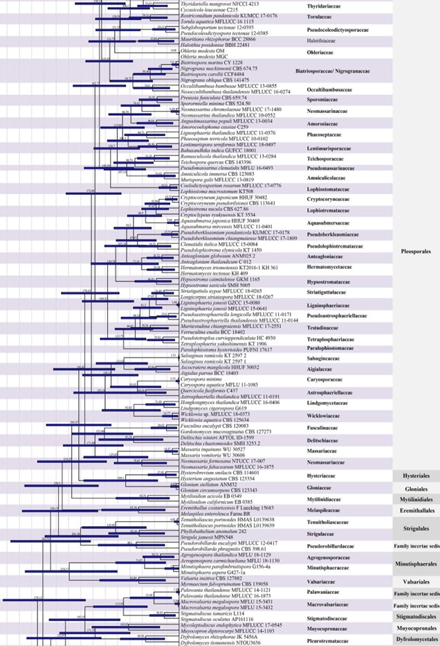
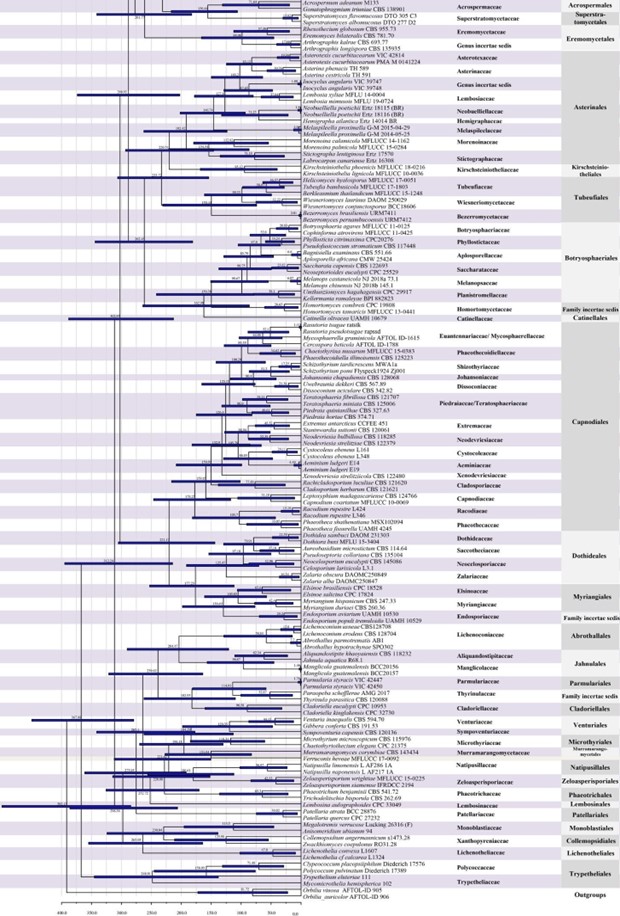
Figure 2 – The maximum clade credibility (MCC) tree of families in Dothideomycetes obtained from a Bayesian approach (BEAST). The fossil minimum age constraints and second calibrations used in this study are marked with green dots. Bars correspond to the 95 % highest posterior density (HPD) intervals. The scale axis shows divergence times as millions of years ago (MYA). Geological periods are indicated at the base of the tree.

Figure 2 – Continued.

Figure 2 – Continued.
Species
Fig. Dacampia hookeri (Material examined: UK, Scotland, Mid Perthshire, on dead mosses on the micaceous soil of Ben Lawers, on thallus of lichen, W.J. Hooker, K(M) 140027, isotype?). a Herbarium label b Specimen of Dacampia hookeri. c Ascomata on white thallus. d Section through ascoma. e Section through peridium. f Asci with pseudoparaphyses. g Young ascus with pale brown, immature ascospores. h-j Mature asci. k-o Ascospores stained by Lactoglycerol. Scale bars: c =1 mm, d =100μm, f =50μm, e, g, h, i, j =20μm, k–o =10μm
