Barria piceae Z.Q. Yuan, Mycotaxon 51: 314 (1994)
Index Fungorum number: IF 362246; MycoBank number: MB 362246; Facesoffungi number: FoF 00031
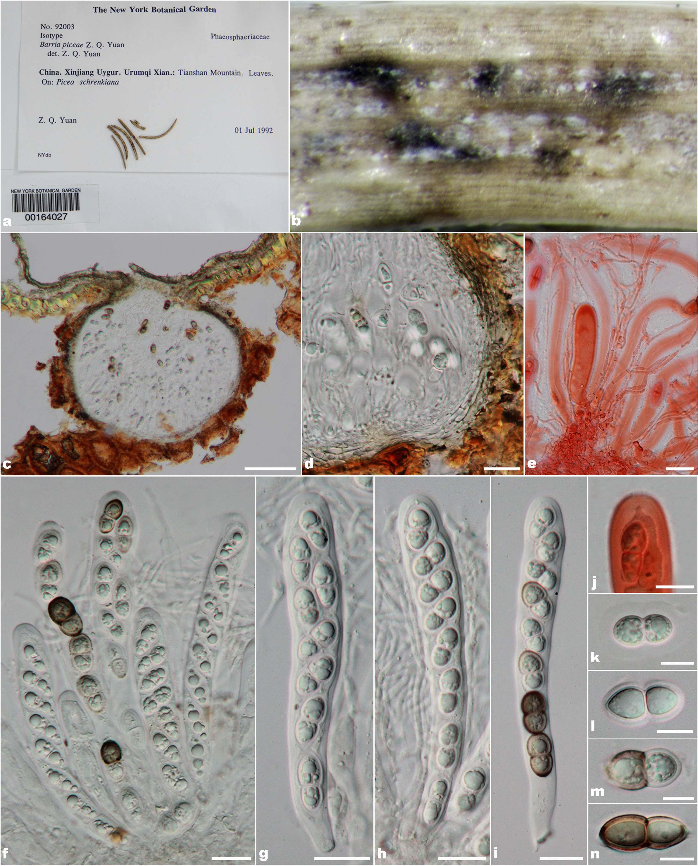
Parasitic on Picea schrenkiana. Ascomata 240–300 µm high, 270–330 µm diam, solitary, sometimes gregarious, immersed, visible as black spots on host surface, uniloculate, globose, brown to dark brown, with centrally opening ostiole. Ostiole apex with or without papilla, ostiolar canal filled with periphyses. Peridium 10–23 µm wide, composed of two cell types, outer layers comprising 3–5 layers, fattened, thin-walled, dark brown to black, pseudoparenchymatous cells of textura angularis or textura prismatica, inner layers composed of 2–3 layers thin-walled cell of textura angularis which are hyaline. Hamathecium composed of numerous, broadly cellular pseudoparaphyses, 1.5–3 µm wide, filiform, branching, anastomosing, hyaline with distinct septate, slightly constricted at the septum, embedded in mucilaginous matrix. Asci (126–)130–170(–185) × (15–)17–20(–23) µm (x̄ = 151.2 × 19 μm, n = 25), 8-spored, bitunicate, fissitunicate, clavate to cylindric-clavate, shortly acute pedicel or sub sessile, apically rounded with well-developed ocular chamber, arising from basal ascoma. Ascospores (18.5–)20–24.5(–27) × 9–12.5 µm (x̄ = 22 × 10.9 μm, n = 30), overlapping, 1–2-seriate, didymospores, ellipsoidal to broadly fusiform, initially hyaline, becoming brown to dark brown at maturity, 1-septate, constricted at the septum, smooth to rough- walled with small guttules, consist of two layers, endospore is thin-walled, with thick-walled at epispore, mostly upper cell larger than the lower cell, surrounded by distinct mucilaginous sheath.
Material examined – China, Xinjiang. Urumqi. Tianshan Mountain., on leaves of Picea schrenkiana, 1 July1992, Z.Q. Yuan_no. 92003, NY 00164027, isotype.
Notes – Barria was introduced by Yuan (1994) and typified with B. piceaewhich and it is monotypic. Barria shares similarities with Didymopleella in the ascomata structure and asci and ascospores characters, but Barria differs from Didymopleella in having textura prismatica and strongly unequal cell of ascospores(Munk 1957, Yuan 1994). Didymosphaeria shows similarities with Barria in having immersed to slightly erumpent ascomata under a clypeus, hyaline pseudoparaphyses, anastomosing frequently above the asci, embedded in mucilage and 1-spetate, ellipsoid ascospores but differ in having a hyaline to pale brown or (rarely) black peridium, consisting of an internal and external layers, flattened or elongated hyphae, textura intricata with trabeculate pseudoparaphyses, distoseptate ascospores (Aptroot 1995), wherein Barria brown to dark brown ascomata comprising several layers of textura angularis to textura prismatica cells in peridium, cellular pseudoparaphyses and asci having well developed ocular chamber bearing ellipsoidal to broadly fusiform, initially hyaline, becoming brown to dark brown at maturity, 1-septate ascospores with distinct mucilaginous sheath. The genus was tentatively accommodated in Phaeosphaeriaceae based on the ascomata and colored and shaped of ascospores (Yuan 1994, Zhang et al. 2012). The phylogenetic investigation is lacking in this genus and thus could not resolve the position in Phaeosphaeriaceae.
In the present study, we propose to exclude this genus from Phaeosphaeriaceae based on morphological characters. Barria associated in gymnosperms, asci clavate with short pedicellate, and didymospores ascospores with thick-walled, while the generic type, Phaeosphaeria associated in angiosperms, asci often broadly cylindrical to cylindric-clavate with sub sessile pedicellate and a phragmospores ascospores. Additionally, most of the genera are confirmed placements in Phaeosphaeriaceae by phylogenetically have phragmospores, scolecospores, or muriform ascospores, but there is not any phylogenetic resolving the didymospores fungi in Phaeosphaeriaceae. However, Barria associated in the same family host as Setomelanomma which the genus was confirmed placement in Phaeosphaeriaceae based on the phylogenetic evidence but they are different by their ascospores type as Barria has didymospores while Setomelanomma has phragmospores ascospores. The ascospores type seems to be effective in distinguishing the genera in Phaeosphaeriaceae. Furthermore, Barria is best fit to genera in Montagnulaceae rather than Phaeosphaeriaceae, thus we tentatively placed Barria in Montagnulaceae but fresh collections, epitypification and molecular work need to be required to prove our classification (Zhang et al. 2012, Hyde et al. 2013).
Fig. 1 – Barria piceae (no. 92003_isotype) a. Herbarium label and specimens of Barria piceae. b. Ascomata on host surface. c. Vertical section through ascomata d. Section through peridium. e. Pseudoparaphyses in congo red reagent. f. Asci with pseudoparaphyses. g-i. Asci. j. Ocular chamber in congo red reagent. k-n. Ellipsoidal to broadly fusiform ascospores. Scale bars: c = 100 µm, d, e f, g, h, i = 20 µm, j-m = 10 µm.

