Asterina gesneraceae (Henn.) Hongsanan & KD Hyde, comb . nov. , Index Fungorum : IF 550716 :
≡ Cocconia gesneraceae Henn., Hedwigia 43: 91 (1904)
≡ Symphaster gesneraceae (Henn.) Theiss. & Syd., Annls mycol. 13(3/4): 217 (1915)
Type: S-F6479.
Epiphytes on surface of leaves. Superficial hyphae with lateral irregular columniform appressoria, which penetrate the host epidermal cells. Sexual state: Thyriothecia 530– 830 diam×49–76 high μm (x = 701.5×64μm, n=6), solitary, scattered, superficial on the leaves, appearing as large circular crusts, easily removed, brown to black, shiny to matt, circular or elliptical. Peridium at the apex and sides composed of dark brown to black thick-walled cells of textura angularis, the lower wall composed of olive brown, poorly defined cells, forming a rather flattened textura angularis, locule composed of light brown cells with asci and pseudoparaphyses arising from that cushion. Hamathecium of up to 1–2μm long, septate, hyaline, branched pseudoparaphyses. Asci 23–29×12–16.5μm (x = 26.5×14.5μm, n=10), 8-spored, bitunicate, globose or subglobose, obovoid to clavate, short pedicellate, thickwalled, apical region of asci usually with a thick opaque region, embedded in mucilage, ascospores released at maturity through an apical fissure above each locule. Ascospores 13–15.5×5.8–6.9μm (x = 14.5×6.2μm, n=10), biseriate, ellipsoid to fusiform, usually two-celled, normally unequal, obviously constricted at septa, upper cell wider, lower cell narrow and longer, hyaline, then turning light greyish-brown to greyish-brown when spores are mature, each cell with oil globules, wall smooth. Asexual state: Unknown.
Material examined: BRAZIL, Rio de Janeiro, Novo Friburgo, on living leaves of Gesneraceae, January 1898, E. Ule 2567 (S-F6479, isotype of Symphaster gesneraceae).
Note: Symphaster is typical to Asterina base on morphology, while difference is only for separate in species levels. This genus is a synonym of Asterina.
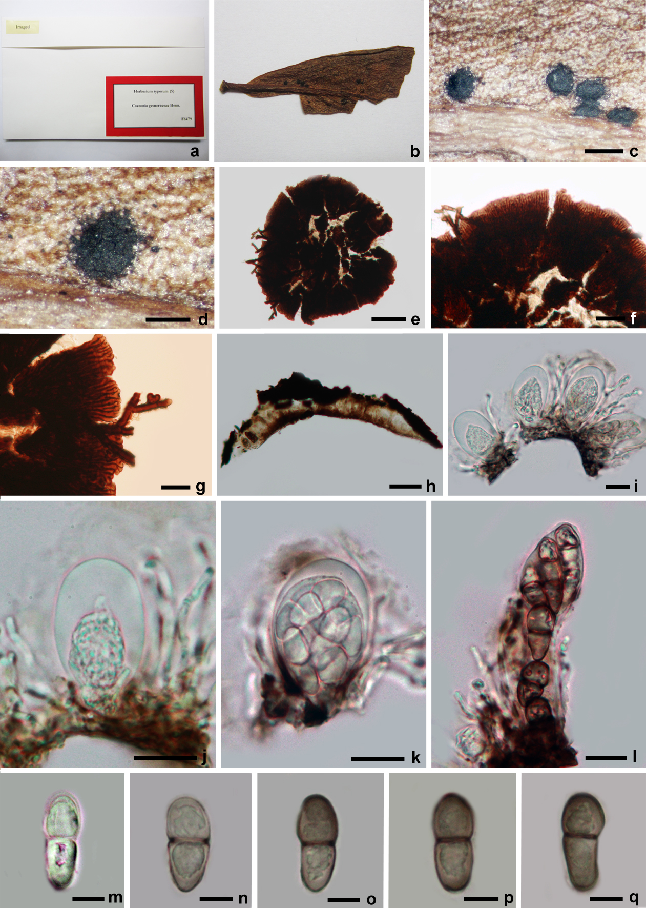
Fig. 1 Asterina gesneraceae (isotype of Symphaster gesneraceae). a Herbarium packet. b Herbarium material. c, d Thyriothecia on the host surface. e–g Squash mount of thyriothecia. Note the superficial hyphae and appressoria. h Hand section of thyriothecia. i–l Asci with ascospores. Note ascus immersed in mucilage. m–q Ascospores. Scale bars: c= 1000μm, d=500μm, e=100μm, f, g=20μm, h=50μm, i–l=10μm, m– q=5μm