Anariste poliothea Syd., Annls mycol. 25(1/2): 76 (1927).
≡ Asterina poliothea (Syd.) F. Stevens, Illinois Biol. Monogr. (Urbana) 17(2): 64 (1939)
Index Fungorum number: IF 274866; Faceoffungi number: FoF 11079
Type: S-F1692, syntype; BPI 690985, syntype.
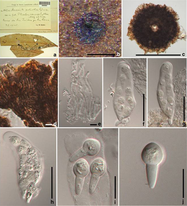
Epiphytes on the upper surface of the leaves. Sexual state: Thyriothecia 238–256 μm diam, solitary, scattered, superficial, easily removed from the host surface, circular, dark brown to black, with ostiole at the centre of the thyriothecium and with bright brown margin. Upper wall composed of interwoven cells, basal wall poorly developed. Hamathecium of hyaline pseudoparaphyses, arranged longitudinally in the thyriothecium. Asci 55–62×11–19μm (x = 54×14.5μm, n=10), 8-spored, cylindric-clavate, fusiform or obclavate, with a knob-like pedicel up to 7μm, apically rounded with an indistinct ocular chamber. Ascospores 16–18×7–9μm (x = 17×8μm, n=10), overlapping 2–3 -seriate, hyaline, 1-septate, weakly constricted at the thin septum, upper cell globose to oval, lower cell conical tapering to the rounded base, wall smooth. Asexual state: Unknown.
Material examined: COSTA RICA, Alajuela, Cerro de San Isidro, near San Ramon, on leaves of Phoebes neurophyllae Mez et Pitt. (Lauraceae), 9 February 1925, H. Sydow (S 1692, syntype); ibid. (BPI 690985, syntype).
Notes: Anariste was established by Sydow (1927) and has remained monotypic until now. Sydow (1927) placed this genus in Microthyriacearum, while von Arx and Müller (1975) and Hosagoudar (2001) placed it in the family Asterinaceae. The genus shares characters of Micropeltidaceae with ostioles at the centre of the thyriothecia, the upper wall composed of interwoven cells, longitudinally arranged asci and cylindrical asci with pedicels. It differs from other genera of Micropeltidaceae in its unusual ascospores. We therefore exclude this genus from the Asterinaceae and place it in the family Micropeltidaceae; molecular data is required to confirm this.
Fig. 1 Anariste poliothea (BPI 690985, syntype). a Material label and specimen. b Appearance of thyriothecium on the host surface. c Squash mount of thyriothecia. d Squash mount of thyriothecium margin. e Pseudoparaphyses. f–h Asci. i, j Ascospores. Scale bars: b=200μm, c= 100μm, d, e=5μm, f–h=20μm, i, j=10μm