Lembosia tenella Lév., Annls Sci. Nat., Bot., sér. 3 3: 58 (1845).
Epiphytes on the lower surface. Superficial hyphae with appressoria. Sexual state: Thyriothecia solitary and scattered, superficial, easily removed from the host surface, black opening by a linear fissure, with basal peridium poorly developed. Upper wall comprising linear, dark cells, which are branched at the margin. Hamathecium comprising vertical asci inclined upwards, pseudoparaphyses not observed. Asci 30–38×21–26μm (x = 36×24μm, n=10), bitunicate, fissitunicate dehiscence not observed, subglobose to ovoid, apedicellate, apical region of asci usually with a thick opaque region, ocular chamber not observed, not staining blue in IKI. Ascospores 15–18×5–7μm (x = 17×5μm, n=10), overlapping, oblong to ovoid, with narrowly rounded ends, hyaline, 1-septate, strongly constricted at the septum. Asexual state: Unknown.
Material examined: NICARAGUA, Herbarium of the U.S. North Pacific Expedition under commanders Ringgold and Rodgers, 1853–1856, collected by C. Wright (PC 0084484).
Notes: Lembosia species are common, widespread and many species are named according to different hosts (Müller and von Arx 1962, Sivanesan and Shivas 2002). Sivanesan and Shivas (2002) recently added three new species of Lembosia from Australia, including L. alyxiae Sivan. & R.G. Shivas, L. araucariae Sivan. & R.G. Shivas and L. syzygii Sivan. & R.G. Shivas, while Hosagoudar (2012) gave an account of the Indian species. We could not locate holotype material of the type species and therefore use a collection from Montagne’s herbarium. We also illustrate a fresh collection of Lembosia from Thailand.
Material examined: THAILAND, Chiang Rai, Mae Fah Luang University, on upper surface of dead leaves, 2013, KD Hyde (MFU 13-0377).
Notes: The morphology of MFU 13-0377 most typical to Lembosia albersii based asci shape and size of ascospores.
Fig. 1 Lembosia tenella (PC 0084484). a, c Appearance of thyriothecia on the host surface. b Squash mount of thyriothecium showing longitudinal fissure. d Vertical section through thyriothecium stained in lactophenol cotton blue. e Asci in Melzer’s reagent. f, g Ascospores within asci, in Melzer’s reagent. Scale bars: b=500μm, c=100μm, d, e=50μm, f=20μm

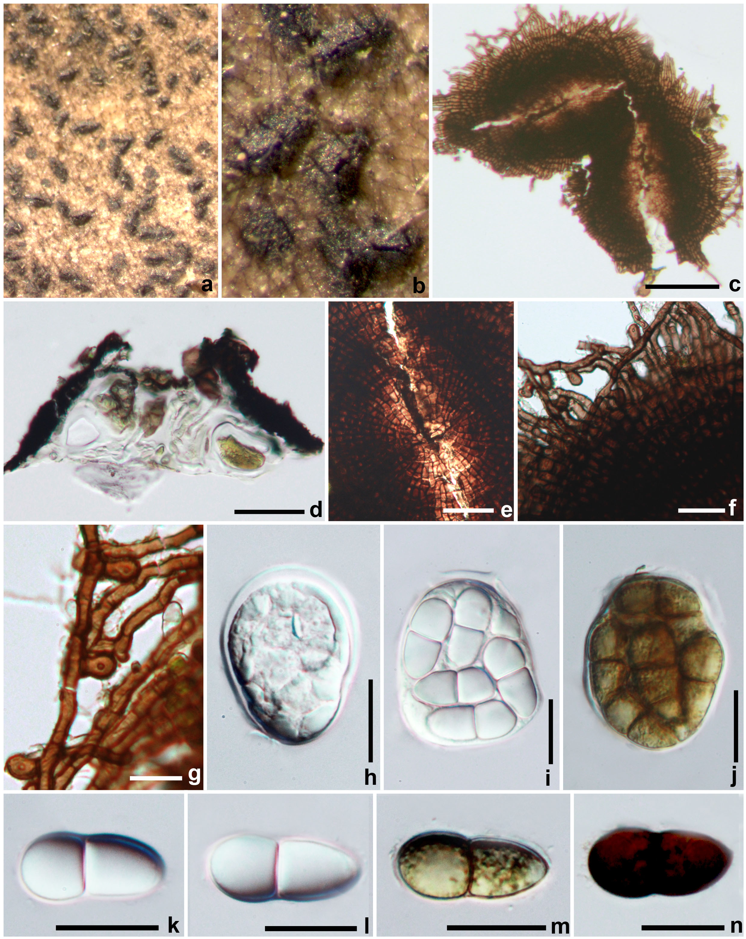
Fig. 2 Lembosia albersii (MFLU 13-0377). a, b Thyriothecia on the host surface. c Squash mount of thyriothecium showing longitudinal fissure. d Section through thyriothecium. f Upper wall of thyriothecium. g Superficial hyphae with appressoria. h, i Immature asci. j Mature asci. k–m Immature ascospores. n Mature ascospores. Scale bars: c=100μm, d=50μm, e, f, h–n=20μm, g=10μm