Anthocanalis sparti Daranagama, E. Camporesi & K.D. Hyde, [as ‘sparti‘], in Daranagama, Camporesi, Tian, Liu, Chamyuang, Stadler & Hyde, Fungal Diversity 73: 211 (2015),
Index Fungorum number: IF 820973; Facesoffungi number: FoF00202
Etymology: In reference to the host, Spartium junceum L.
Holotype – MFLU 14–0225
Saprobic on stems, branches and twigs. Sexual morph: Ascomata immersed, solitary and scattered, appearing as raised black spots, in cross section 280–300 µm diam. and 250–265 µm high (x̄ = 293 × 256 µm, n = 10), conical to globose, with poorly developed clypeus and shiny papilla. Peridium 30–35 µm (x̄ = 31 µm, n = 10) diam. at base, comprising two cell layers, outwardly comprising thick-walled, light brown cells of textura irregularis and inwardly comprising thin-walled, hyaline cells of textura angularis. Paraphyses 5 µm diam. (x̄ = 5 µm, n = 30), slightly longer than the asci, numerous, filamentous, septate. Asci 110–150 × 5–8 µm (x̄ = 125 × 6 µm, n = 30), 8-spored, unitunicate, cylindrical, short-pedicellate, apically rounded with a rectangular to discoid, J+, apical apparatus, 1.8–2.5 µm high × 0.5–1 µm diam. (x̄ = 2.3 × 0.7 µm, n = 20). Ascospores 14–18 × 6–8.5 µm (x̄ = 15.5 × 7.3 µm, n = 40), overlapping uniseriate, limoniform, with one side flattened to concave, dark brown, smooth-walled, mucilaginous sheath clearly visible at the flattened side, sheath 1–2 µm thick, constricted at centre along the flattened edge forming an indentation, germ slit, full length, straight. Asexual morph: Conidiophores up to 50–80 µm long and 1.5–2 µm diam (x̄ = 1.6 µm, n = 40), complex, nodulisporium-like, hyaline, branched. Conidiogenous cells 10–12 µm long and 1.5–1.8 µm diam. (x̄ = 11.5 × 1.6 µm, n = 40), terminal at the branches, cylindrical. Conidia 5–5.5 × 2–2.5 µm (x̄ = 5.3 × 2.3 µm, n = 40), hyaline, ellipsoidal, aseptate, unicellular.
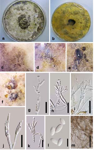
Culture characteristics – Colonies on Difco OA at 25–27 oC reaching the edge of 9 cm Petri-dish in 4 weeks, at first whitish, felty, azonate, with diffuse margins, developing sporulating regions as black spots in the centre and periphery of the culture; reverse turning Citrine (13) or Isebellien (65). Producing dense conidiophores.
Material examined – ITALY, Province of Forlì-Cesena, Corniolo-Santa Sofia, on Spartium junceum L. (Fabaceae), 25 October 2012, E. Camporesi IT848 (MFLU 14–0225, holotype), ex-type culture, MFLUCC 14–0010 = ICMP; ibid, 25 October 2012, E. Camporesi IT848-bis (PDD, isotype); Monte Riccio, Bagno di Romagna, 20 December 2013, E. Camporesi IT848–tris (MFLU 14–0226, PDD paratype), ex-paratype culture, MFLUCC 14–0557.
Notes – Anthocanalis sparti is reminiscent to Anthostomella colligata K.D. Hyde & B.S. Lu, A. flagellariae (Rehm) B.S. Lu & K.D. Hyde and A. francisiae K.D. Hyde and these taxa may need transferring to Anthocanalis following molecular study. Anthostomella colligata has deeply immersed ascomata, a well-developed clypeus and long neck, a peridium consisting of cells with light brown walls at the inside and dark brown, thick walls, towards the outside, and ascospores with thick mucilaginous sheath, which is constricted in the centre at both edges of the ascospores. Anthostomella flagellariae has immersed ascomata, with peridium comprising of brown cells, relatively short-pedicellate asci and ascospores with a mucilaginous sheath, which is wider at each end. Anthostomella francisiae is visible on the host as oval regions with a disc–like clypeus made up of host and fungal hyphae, asci with a cylindrical, J+, apical apparatus and ascospores, with pad-like appendages at each end (Lu and Hyde 2000b). In contrast, Anthocanalis sparti has immersed ascomata appearing as globose to conical, black spots, with a poorly developed clypeus and shiny papilla, a peridium with hyaline cells of textura angularis and light brown cells of textura irregularis, asci with J+ discoid to rectangular, apical apparatus and uniseriately arranged ascospores with a prominent sheath only at the ventral side of the ascospore and straight, full-length germ slit.
Fig. 1 Anthocanalis spartium (MFLU 14–225, holotype). a Habitat. b Ascomata in wood. c Cross section of an ascoma. d peridium. e Asci in Meltzer’s reagent with J+ rectangular shaped apical apparatus and straight spore-length germ slit (in mature ascospores). f Mature asci in water. g–j Ascospores in water with mucilaginous sheath. Scale bars: a = 500 µm, b = 1 mm c, d=50 µm, e–j = 10µm
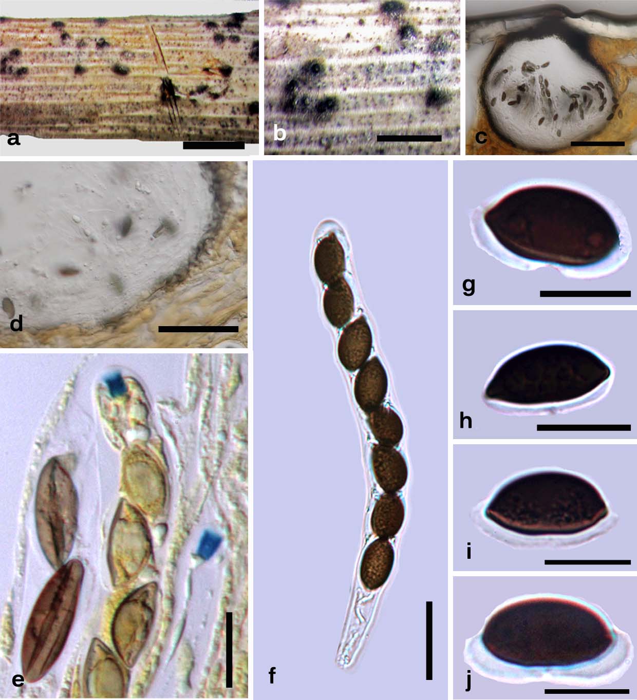
Fig. 2 Anthocanalis sparti (MFLU 14-0225, holotype). a, b Colony on OA after 4 weeks a From above. b From below. c–e Development of sporulating regions after 7, 14 and 21 days. f Oil droplet. g–i Conidiogenous structures. j, k Conidiogenous cells. l Conidiam. m Mycelia. Scale bars: g–k=20 μm, l = 5 μm, m = 30 μm.