Setomelanomma holmii M. Morelet, Bull. Soc. Sci. nat. Arch. Toulon et du Var 36(no. 227): 15 (1980).
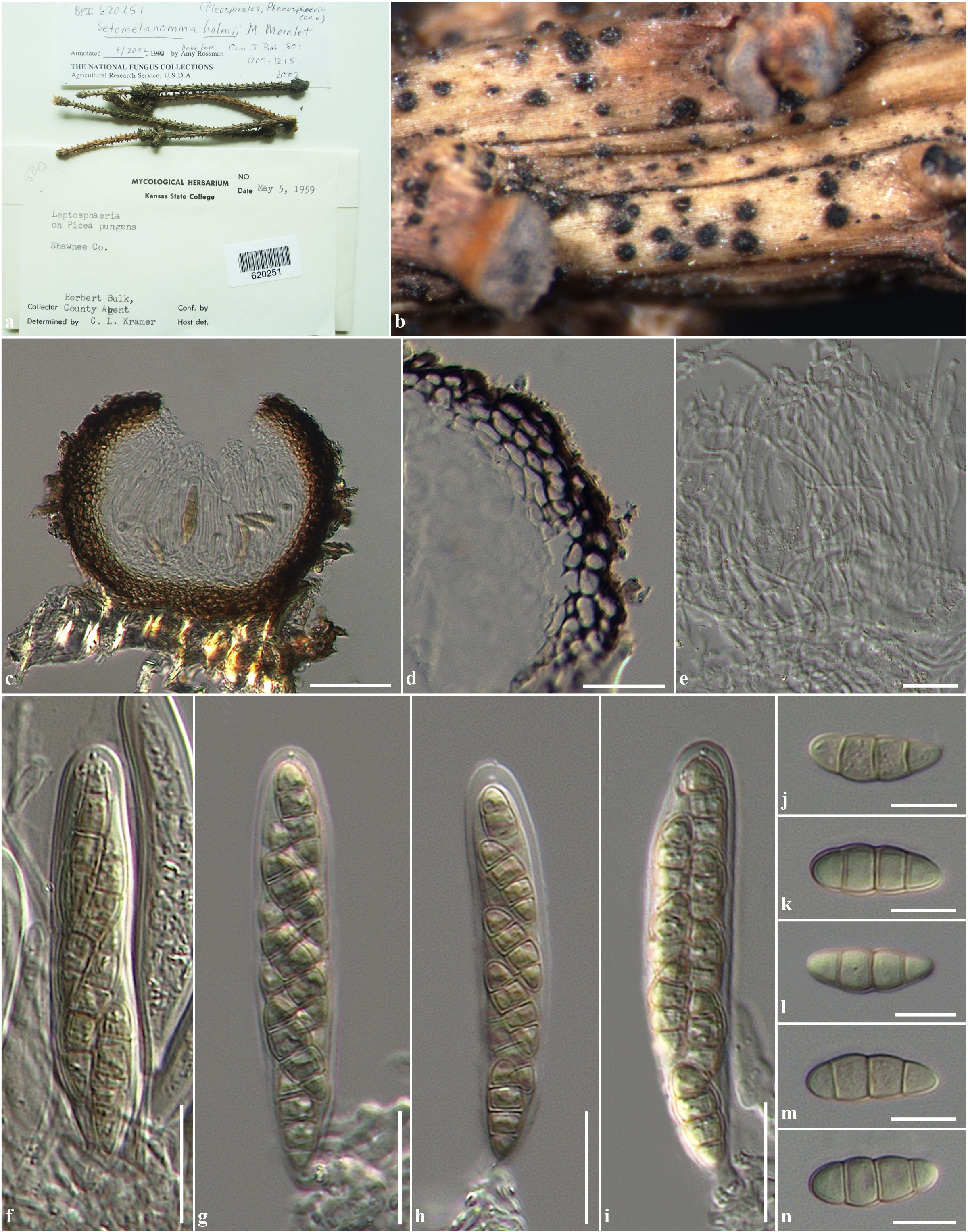
Pathogenic on branches of Picea. Sexual state: Ascomata 100–150 μm high, 140–175 μm diam., scattered, gregarious semi-immersed to erumpent through host tissue, becoming superficial, visible as abundant, small black dots on the host surface, uniloculate, globose to subglobose, covered by setae, dark brown to black, with central ostiole. Peridium 12.5–26 µm wide, thin-walled, of equal thickness, composed of 5–7 layers of black, broadly pseudoparenchymatous cells, arranged in a textura angularis to textura globulosa. Hamathecium composed of numerous, 1.5–3 µm wide, filamentous, frequently anastomosing, broad cellular pseudoparaphyses, with distinct septa, embedded in mucilaginous matrix. Asci (67–)68–77(–85) × (13–)15–17(–20) µm ( = 75.2 × 15.2 μm, n = 20), 8-spored, bitunicate, fissitunicate, broadly cylindrical to cylindric-clavate, subsessile to short pedicellate, apically rounded with well-developed ocular chamber. Ascospores (18–)21–23(–24) × (6–)7–9(–11) µm ( = 21.3 × 8.2 μm, n = 20), overlapping 1–2-seriate, phragmosporous, ellipsoidal to broadly fusiform, initially hyaline to pale yellowish, becoming light brown to brown or yellowish-brown at maturity, initially forming one median septum, becoming 3-septate at maturity, slightly curved, often constricted at the median septum, smooth and thick-walled. Asexual state: see under Xenoseptoria neosaccardoi Quaedvlieg et al. (2013)
Material examined: USA, Kansas, Shawnee Coounty, on branches of Picea pungens Engelm (Pinaceae), 5 May 1959, B. Herbert (BPI 620251).
Fig. 1 Setomelanomma holmii (BPI 620251) a Herbarium label and specimens. b Ascomata on substrate. c Section through ascoma. d Section through peridium. e Cellular pseudoparaphyses. f-i Asci. j-n Ascospores. Scale bars: c = 50 µm, d, e, f, g, h, i = 20 µm, j, k, l, m, n = 10 µm.