Pleospora herbarum (Pers.) Rabenh., Klotzschii Herb. Viv. Mycol. 2: no. 547 (1854).
≡ Sphaeria herbarum Pers., Syn. meth. fung. (Göttingen) 1: 78 (1801)
Index Fungorum number: IF 208023; Facesoffungi number: FoF 00535
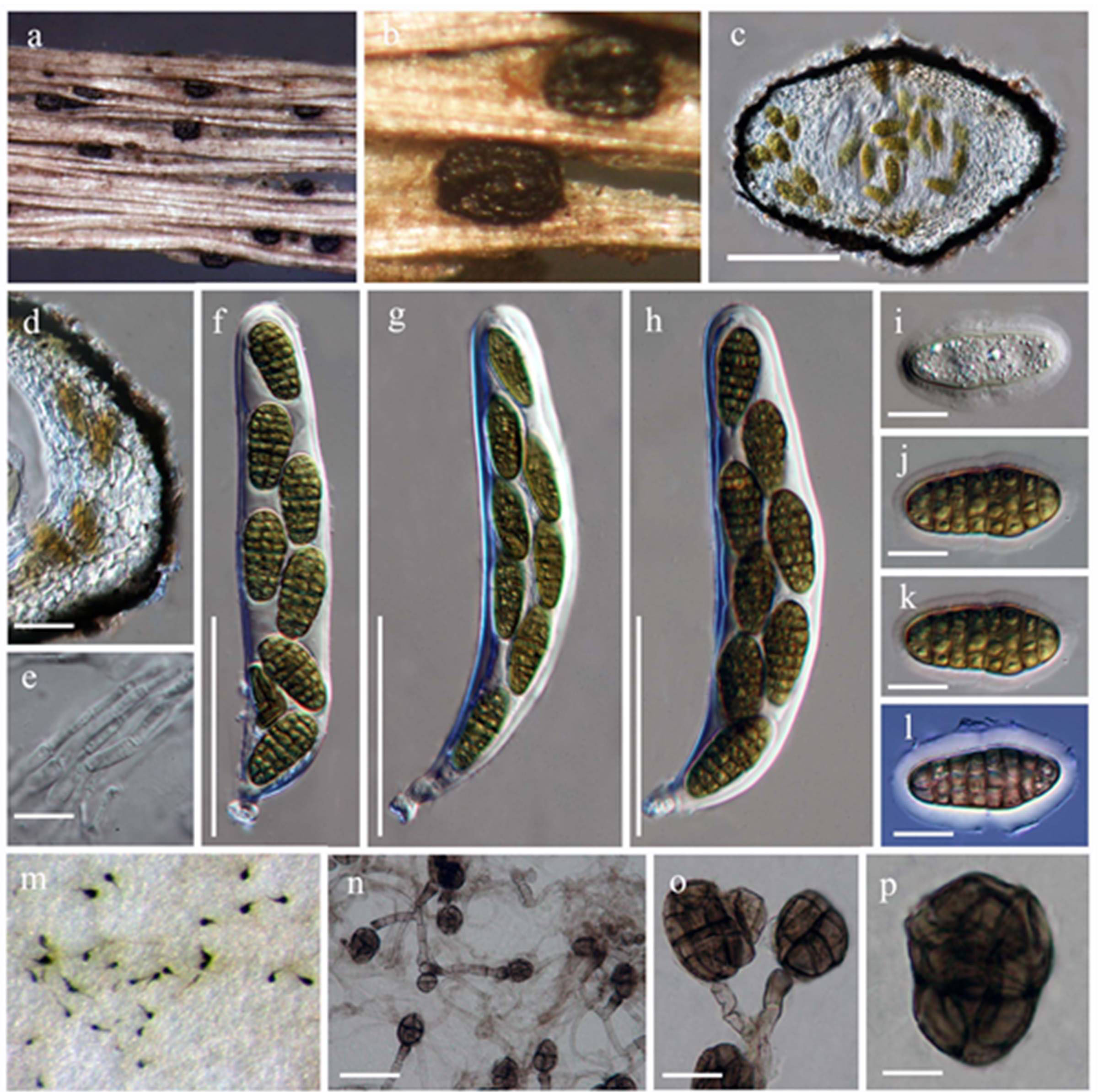
Habitat terrestrial, marine, saprobic or parasitic. Sexual morph: Ascomata 110–240 × 250–460 μm small to medium-sized, immersed, erumpent to superficial, base not easy to remove from the substrate, broadly to narrowly oblong and flattened, ostiolate. Ostiole papillate, black, smooth, ostiolar canal filled with hyaline cells. Peridium 30–50 μm ( = 42 μm, n=10) thin, usually with two layers, thick at the sides and thinner at the base, outer layer of heavily pigmented thick-walled cells of textura angularis, inner layer composed of hyaline thin-walled cells of textura angularis, coriaceous. Hamathecium of 2–3 μm ( = 2.5 μm, n=10) cellular, hyaline, septate, broad, dense pseudoparaphyses. Asci 100–225 × 25–30 µm ( = 142 × 28 µm, n=20), 8-spored, bitunicate, fissitunicate, cylindrical to clavate, with furcate pedicel and minute ocular chamber. Ascospores 25–40 × 12–15 µm ( = 33 × 14 µm, n=40), uniseriate or partially overlapping, mostly ellipsoidal, muriform, brown or pale brown, with or without sheath. Asexual morph: Stemphylium. Conidiophores 4–6 μm ( = 5 μm, n=10) macronematous, mononematous, scattered or caespitose, unbranched or rarely loosely branched, straight or flexuous, usually nodose with a number of vesicular swellings, pale to mid brown or olivaceous brown, smooth or in part verruculose. Conidiogenous cells monoblastic, integrated, terminal, percurrent, at first clavate or subsphearical with the wall at the apex thin. Conidia 16–22 × 11–18 µm ( = 21 × 16 µm, n=20) solitary, dry, acrogenous, oblong, rounded at the ends, ellipsoidal, obclavate or subspherical, pale to mid-dark or olivaceous-brown, smooth, verrucose or echinulate, muriform, often constricted at one or more of the septa, cicatrized at the base. Colonies effuse, grey, brown, olivaceous-brown or black, velvety or cottony.
Materials examined – ITALY, Forlì-Cesena, Montevescovo, on the dead stem of Brassica nigra (Brassicaceae), 01 January 2012, E. Camporesi IT 82 (MFLU 12-2216) – living culture (MFLUCC 14-0261); ITALY, Tessello – Cesena, on the dead stem of Cirsium sp (Asteraceae), 16 December 2013, E. Camporesi IT 956 (MFLU 14-0762) – living culture (MFLUCC 13-0344); ITALY, Massera – Predappio, on dead fruits of Lunaria rediviva (Brassicaceae), 16 December 2013, E. Camporesi IT 958 (MFLU 14-0763) – living culture (MFLUCC 13-0266, ICMP)
Notes – Pleospora was originally described by Rabenhorst (1857) and is typified by Pleospora herbarum (Pers.) Rabenh. (Kirk et al. 2008). Initially, the genus was included in Sphaeriales and later Pseudosphaeriales and Pleosporales, respectively (Wehmeyer 1961). All species belonging to Pleospora have muriform ascospores (Wehmeyer 1961, 1975). Pseudoparaphyses arrangement (downward growing) within the ascomata of “Pleospora-type” development (Luttrell 1951) is considered to be the main feature of the genus. Various authors have included and excluded different species in Pleospora at various times. Barr (1981) placed Curreya in Pleospora however, von Arx and van der Aa (1983) treated it as a separate genus because of its Coniothyrium asexual morph. Petrak (1952) transferred Graphyllium to Pleospora and noted that the elongated ascomata and closely grouped rows of small ascomata are not sufficient to recognize the genus. Barr (1987b, 1990b) supported this proposal. Due to the heterogenous nature of Pleospora, several subgenera have been included. i.e. Teichosporoides contains species of Pleospora with immersed ascomata and Pleosphaeria with superficial and setose ascomata (Wehmeyer 1961). Pleospora species have a wide host range, especially on monocotyledons as well as dicotyledonous plants (Wehmeyer 1975).
There are numerous species of Dothideomycetes that have muriform ascospores and have at one time or another been placed in Pleospora (Wehmeyer 1961). Pleospora is, however, rather distinctive and should be confined to species with characters that are similar to Pleospora herbarum. The ascomata in P. herbarum are immersed and usually become erumpent, the peridium is unusual in having three strata, a thin inner layer of thin-walled, hyaline to light brown flattened cells, a relatively wide central layer of thin-walled, hyaline to light brown angular cells, and an outer very thin black amorphous cells, which gives the blackened colour to the ascomata. The asci are broadly clavate and have a distinctive, squarish, wide ocular chamber and ascospores are muriform with at least three longitudinal septa per transverse row and surrounded by a mucilaginous sheath. The asexual morph is Stemphylium (Zhang et al. 2012; Hyde et al. 2013). For this reason, several Pleospora-like species are now transferred to other genera (e.g. Curreya).
Based on our phylogenetic assessment we proposed to confine Pleospora to one well-supported clade based on the type strains of P. herbarum (CBS 191.86) along with the other alternative strains of P. herbarum used in this study (P. herbarum MFLUCC 13-0344, P. herbarum MFLUCC 13-0266 and P. herbarum (MFLUCC 14-0261), sister to Paradendryphiella clade applying GCPSR (Taylor et al. 2000; Dettman et al. 2003). By implementing this, we treat some putative strains, such as P. tomatonis (CBS109844), P. papaveracea (CBS432.50), P. tarda (CBS 714.68) and P. halophila (CBS410.73) as alternative strains of P. herbarum. This treatment leads to clarification of the paraphyletic nature of Pleospora within the family Pleosporaceae.
Fig. 1 Pleospora herbarum (MFLU12-2216). a. Ascomata on host substrate. b. Close up of ascoma .c . Section of ascoma. d. Close up of the peridium. e. Cellular, hyaline, septate, broad pseudoparaphyses. f-h. Asci with short, broad pedicel bearing 8 ascospores i-l. Mature and immature ascospores with mucilaginous sheath. Stemphylium sp. m-p. Conidiophores with pale to mid dark or olivaceous brown, smooth conidia. Scale bars: c = 100 μm, d = 50 μm, e = 20 μm, f-g = 60 μm, i-k = 10 μm, l-o = 10μm.