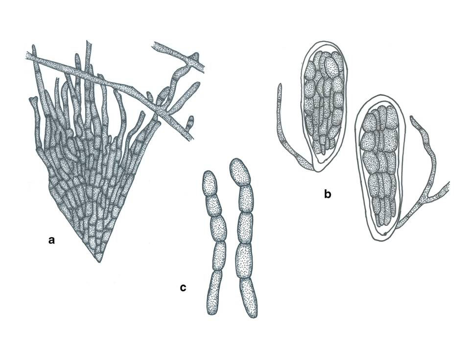
Parasterinopsis sersalisiae (Hansf.) Bat., Atas Inst. Micol. Univ. Recife 1: 327 (1960).
≡ Patouillardina sersalisiae Hansf., Proc. Linn. Soc. London 156: 117 (1944)
Epiphytes on the lower surface of Synsepalum cerasiferum. Superficial hyphae abundant, superficial, light brown, septate, branching and spreading over the host surface. Appressoria 6– 12.5×3–5μm, 2-celled, lateral, narrowly ovate. Sexual state: Thyriothecia 170–250μm diam, superficial, scattered, black, opening by a huge irregular fissure when wet, with basal peridium poorly developed. Upper wall composed of dark to brown filaments at the outer rim, which radiate towards the rim, where they are slightly lighter. Hamathecium of 2– 2.5μm, filamentous, branched pseudoparaphyses and vertically arranged asci. Asci 48–60×48–53μm (x = 50×15μm, n=5), 8-spored, bitunicate, oblong, apedicellate, lacking an ocular chamber, not staining blue in IKI. Ascospores 25–32× 8–11μm (x = 29×10μm, n=5), fasciculate, cylindrical, hyaline to brown, 1–4-septate, constricted at the septa, with upper cell larger and broadly rounded. Asexual state: Unknown.
Material examined: UGANDA, Entebbe, on leaves of Synsepalum cerasiferum (Welw.) T.D. Penn. (as Sersalisia edulis S. Moore) (Sapotaceae), C.G. Hansford Typus no 3650, Herb Mycologist Dept Agriculture Uganda, December 1944 (URM 11923, exs. 6979, isotype).
Notes: The ascospores in the specimen were not clear and therefore we illustrate whatmay be a broken ascospore and we also redraw the ascospores from the protologue (fig 2).
Fig. 1 Parasterinopsis sersalisiae (isotype). a Herbarium specimen. b Thyriothecia and hyphae on the lower surface of the host. c, d Squash mount of thyriothecia in Melzer’s reagent. e Vertical section through thyriothecium in cotton blue reagent. f Asci in cotton blue reagent. g Asci and ascospores in cotton blue reagent. Scale bars: b=0.5 mm, c–e= 100μm, f, g=10μm
Fig. 2 Parasterinopsis sersalisiae (redrawn form Batista 1960b). a Thyriothecia margin and hyphae on the lower surface of the host. b Asci with pseudoparaphyses. c Ascospores


