Marielliottia biseptata (Sacc. & Roum.) Shoemaker, Can. J. Bot. 76(9): 1560 (1999)
≡ Helminthosporium biseptatum Sacc. & Roum., Revue Mycol. (Toulouse): 56 (1881).
≡ Brachysporium biseptatum (Sacc. & Roum.) Sacc., Sylloge Fungorum 4: 428 (1886).
≡ Drechslera biseptata (Sacc. & Roum.) M.J. Richardson & E.M. Fraser, Transactions of the British Mycological Society 51 (1): 148 (1968).
≡ Pyrenophora biseptata (Sacc. & Roum.) Crous, CBS Biodiversity Series 13: 257 (2013)
= Helminthosporium biforme E.W. Mason & S. Hughes, Transactions of the British Mycological Society 30: 114 (1948).
= Drechslera biforme (E.W. Mason & S. Hughes) Subram. & B.L. Jain (1966).
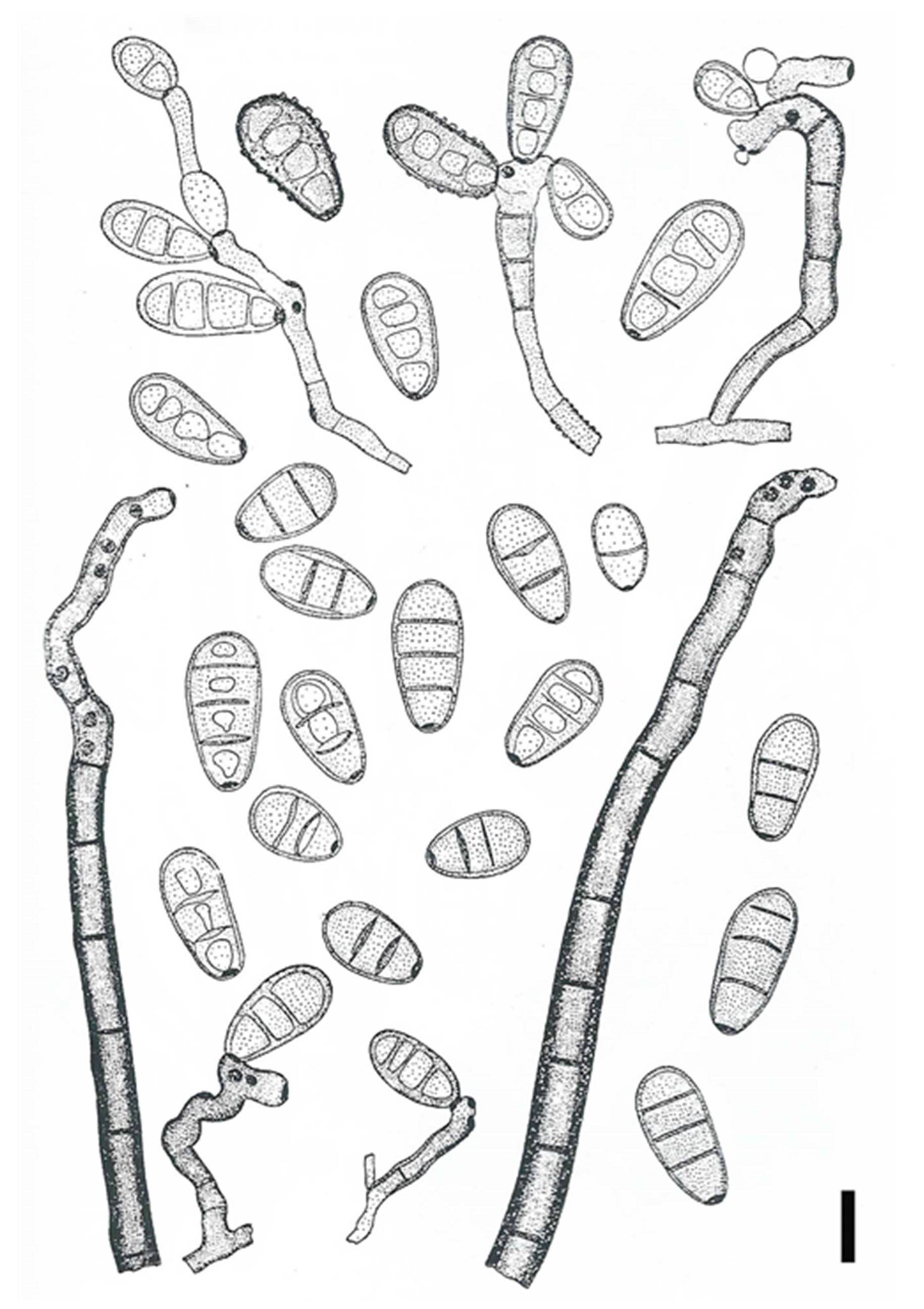
Saprobic on stems. Sexual morph: unknown. Asexual morph: Conidiophores two types. Macronematous conidiophores from stroma, single to many in a cluster, erect to divergent, long, broad at base, gradually narrowed towards apex, closely septate at intervals of 10–18 mm, dark chocolate brown; sporogenous area gnarled, frequently bent, with numerous dark brown scars, apex truncate, bearing an “ear” of conidia, 100–800 × 10–15 µm (at base), 9–11 µm wide near middle, 6–8 µm wide near apex. Conidiogenous cells tretic, integrated, terminal, sympodially proliferating, cylindrical, cicatrized, stromata shallow ellipsoidal to depressed globose, 100–250 × 100–150 µm. Stroma initials cigar shaped, within host epidermal cells, subdivided into cubical cells. Micronematous conidiophores arising from hyaline to pale yellow hyphae (in culture), slender at base, sparingly septate, short, erect, sporogenous area gnarled, densely scarred, dark chocolate brown, usually simple, rarely branched, 15–105 × 5– 8 µm. Conidiogenous cells tretic, integrated, terminal, sympodially proliferating, cylindrical, cicatrized. Conidia obovoid, 3-septate, uppermost septum inconspicuous or lacking in some, rarely constricted at septa, thick walled, light to dark olive brown except lighter basal cell, narrowed to base, bearing a broad black nonprotruding scar 3–4.5 µm wide, (21–)32–45(–49) × (9–)11–13(–17) µm. Germination by one or two (three) oblique tubes near scar, rarely one hypha from apex and very rarely through the scar (Shoemaker 1998).
Notes: Marielliottia was introduced by Shoemaker (1998) to accommodate three species formerly treated in Drechslera namely D. biseptata (Sacc. & Roum.) M.J. Richardson & E.M. Fraser, D. dematioidea (Bubák & Wróbl.) Scharif and D. triseptata (Drechsler) Subram. & B.L. Jain. The genus is typified with Marielliottia biseptata. The species differ from Drechslera in having mostly three-septate, obovoid to ovoid conidia that germinate primarily from the basal cell, occasionally from the apical cell, and not from the central cells (Shoemaker 1998). These fungi are occasional parasites of cereals and grasses, are sometimes seed borne, and may infect non-grass plants (Shoemaker 1998).
Currently no sequence data available for Marielliottia species in GenBank. Therefore, fresh collections of the type species of the genus are needed so that molecular data can be obtained to verify the natural taxonomic affinities of this genus. Based on the morphological similarities with Drechslera, we tentatively classified Marielliottia in Pleosporaceae.
Fig. 1 Marielliottia biseptata (Redrawn from Drechslera biseptata in Ellis (1971), Fig 288.). Scale bars: 650 μm