Inocybe brunneosquamulosa K.P.D. Latha & Manim.
MycoBank number: MB 816735 Facesoffungi number: FoF: 2176
Etymology: referring to the brown squamules on the pileus surface.
Holotype: CAL 1308
Basidiocarps small. Pileus 7–11 mm diam., convex with a small umbo when young, becoming broadly convex still with a small umbo at maturity; surface initially dark brown (6F8/OAC636) on the squamules and brownish-orange (6C4/OAC653) elsewhere, becoming dark brown (6F7/OAC637) on the squamules, greyish-orange (6B3/OAC633) on the fibrils and brownish-grey (6C2/OAC662) elsewhere at maturity, appressed- to recurved-squamulose all over when young, becoming appressed- to recurved squamulose on and around the umbo and appressed-fibrillose and rimose towards the margin; margin incurved, becoming decurved, crenate or wavy. Lamellae sinuate, close, initially orange grey (6B2/OAC634), becoming brownish-orange (6C4/OAC655) or light brown (6D4/OAC686) at maturity, up to 1.5 mm wide, with lamellulae of 1 length; edges fimbriate, whitish. Stipe 17–19 × 1–2 mm, central, terete, equal, cartilaginous, solid; surface brownish-orange (6C3/OAC633) all over, appressed-fibrillose in most parts, slightly-recurved fibrillose and finely pruinose towards the apex; base not enlarged. Odour and taste not distinctive. Basidiospores 8–10 × 5–6.5 (9 ± 0.6 × 5.9 ± 0.4) µm, Q = 1.3–1.9, Qm = 1.5, smooth, ellipsoid to subphaseoliform, slightly thick-walled, yellowish-brown. Basidia 21–30 × 11–13 µm, clavate, thin-walled, hyaline, 4-spored; sterigmata up to 4.5 µm long. Pleurocystidia absent. Lamella-edge heterogeneous. Cheilocystidia 17–39 × 11–18 µm, versiform: clavate, utriform, fusiform, cylindrical with an obtuse apex, occasionally subglobose or rarely pedicellate or septate, hyaline with faint hyaline encrustations, thin- to slightly thick-walled. Lamellar trama subregular; hyphae 6–15 µm wide, thin-walled, hyaline or pale yellow. Subhymenium pseudoparenchymatous. Pileus trama subregular, composed of both narrow and inflated hyphae; hyphae 3–30 µm wide, pale yellow, thin-walled. Pileipellis a cutis frequently disrupted with trichodermal patches, often a perfect trichoderm at the centre; hyphae 8–16 µm wide, thin- to slightly thick-walled, with a brown wall pigment and dense, yellowish-brown or brown spiral encrustations; terminal cells 25–52 × 7–11 µm, clavate or cylindrical with an obtuse apex, thin- to slightly thick-walled. Stipitipellis a cutis often disrupted by loose hyphal projections scattered over the entire surface of the stipe and with bunches of caulocystidia confined to the extreme stipe apex; hyphae 5–11 µm wide, thin- to slightly thick-walled, with a pale yellowish-brown wall pigment and faint hyaline encrustations; terminal cells, 21–49 × 5–7 µm, cylindrical or flexuous-cylindric, slightly thick-walled, with a pale yellowish-brown wall pigment and faint hyaline encrustations. Caulocystidia 22–50 × 11–14 µm, catenulate, clavate, inflated clavate or obovoid, rarely septate, hyaline, occasionally with faint, hyaline encrustations, thin- to slightly thick-walled. Stipe trama hyphae with dense, yellowish-brown oleaginous contents. Clamp connections seen on all hyphae.
Habitat: on the ground, scattered around Vateria indica (Dipterocarpaceae) trees.
Specimen examined: INDIA, Kerala State, Ernakulam District, Kochi, Thevakkal, Ponnakkudam Kavu sacred grove, 25 August 2014, K.P.D Latha DKP264 (CAL 1308, holotype). GenBank numbers ITS: KX073582; LSU: KX073586; RPB2: KX073589.
Notes: Small basidiocarps with a dark brown, squamulose and fibrillose-rimose pileus; a fibrillose stipe with a finely pruinose apex and an abruptly ending base; smooth, ellipsoid to subphaseoliform basidiospores; a hymenium devoid of pleurocystidia; versiform cheilocystidia
with occasional, faint, hyaline encrustations; a cutis-type pileipellis which is disrupted with trichodermal patches and a cutis-type stipitipellis disrupted by loose, hyphal projections and often with caulocystidia at the extreme stipe apex are the salient features of I. brunneosquamulosa. Inocybe fuscospinulosa, a species originally described from Indonesia (Horak 1980b) and also reported from Sri Lanka (Pegler 1986), seems to be somewhat similar to I. brunneosquamulosa in having a pileus of rather similar colour and surface features, a fimbriate lamella-edge, a fibrillose stipe, a hymenium devoid of pleurocystidia, a trichoderm-type pileipellis and the presence of cheilocystidia. Inocybe fuscospinulosa, however, is distinguished from the I. brunneosquamulosa in having larger basidiocarps with a densely squamulose pileus, crowded, adnexed, tobacco brown lamellae, a reddish-brown tinted stipe with occasional scales, smaller (6.5–8 × 4–5 µm) and ovoid basidiospores, cylindric to subfusoid cheilocystidia devoid of encrustations and a stipitipellis lacking caulocystidia. Inocybe brunneosquamulosa is also somewhat similar to I. umbrinovirens E. Horak, a species so far known only from Papua New Guinea (Horak 1980b), in having a somewhat similar-coloured pileus with almost similar surface features, a fibrillose stipe, smooth basidiospores, a hymenium devoid of pleurocystidia, the presence of cheilocystidia with encrusting pigment and a trichoderm-type pileipellis. However, the characters such as the larger basidiocarps with differently-shaped pileus, chocolate brown, adnexed, crowded lamellae, a hollow stipe with a greenish base, larger (10–12.5 × 7–8.5 µm) and ovoid basidiospores, larger basidia, cheilocystidia that are terminal elements of lamellar trama, the absence of caulocystidia and a strong odour make I. umbrinovirens different from I. brunneosquamulosa.
Inocybe squamata J.E. Lange, a species widespread in Europe and USA (Cripps 1997) and also reported from Kerala (Pradeep and Vrinda 2010; Mohanan 2011) is similar to I. brunneosquamulosa in having a pileus of somewhat similar texture, rather similarly attached lamellae, a fibrillose stipe, subphaseoliform basidiospores, a hymenium devoid of pleurocystidia, the presence of cheilo- and caulocystidia and similar type of pileipellis. However, I. squamata has larger basidiocarps with a differently-coloured pileus, thick, broad, yellow brown lamellae, a longer, white stipe, larger basidiospores (9–11.5 (13) × (5) 5.5–6.5 µm), occasional 2-spored basidia and narrowly clavate cheilo- and caulocystidia lacking encrustations.
Comparison of the ITS (679 bp), LSU (935 bp) and RPB2 (699 bp) sequences derived from I. brunneosquamulosa with the nucleotide sequences available in GenBank numbers showed that I. brunneosquamulosa has distinct sequences. Inocybe species MCA562 resulted as the closest hit in megablast searches with ITS (GenBank numbers JQ408785; Identities = 630/682 (92%)), LSU (GenBank numbers JN975016; Identities = 921/936 (98%)) and RPB2 (GenBank numbers JQ421077; Identities = 673/699 (96%)) sequences. Inocybe species MCA562 is a collection from Japan, but its morphological features are unavailable for comparison as it remains unpublished.
The phylogenetic placement of I. brunneosquamulosa is shown in the phylogram generated from the ML analysis of RPB2 sequence data matrix. In the ML analysis, I. brunneosquamulosa nested in the Pseudosperma clade with maximum support (100% ML) where it paired with Inocybe species MCA562 and had a strong support (96% ML).
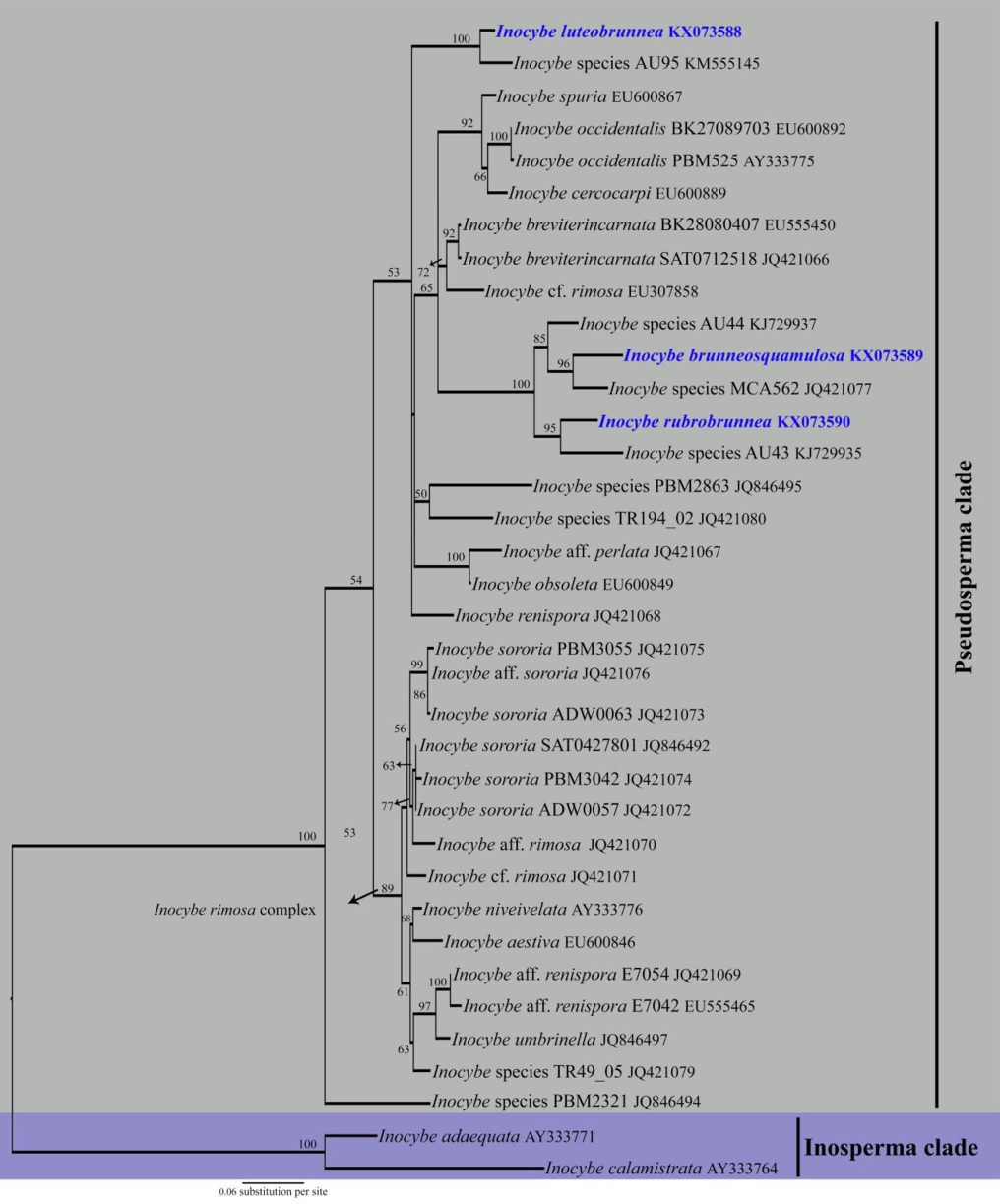
Phylogram generated from maximum likelihood (RAxML) analysis based on RPB2 sequence data matrix for 34 Inocybe species. Sequences of Inocybe species belonging to the Pseudosperma clade used in this study have been selected from a previous analysis of Kropp et al. (2013). Values at nodes indicate bootstrap support. BS values ≥50% are shown. Inocybe luteobrunnea, I. brunneosquamulosa and I. rubrobrunnea are in pink to highlight its phylogenetic position in the tree. The tree is rooted with I. adaequata and I. calamistrata of Inosperma clade.
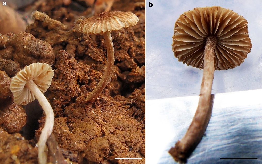
Inocybe brunneosquamulosa (CAL 1308, holotype). a, b Basidiocarps. Scale bars: a, b = 5 mm (photos by K. P. Deepna Latha)
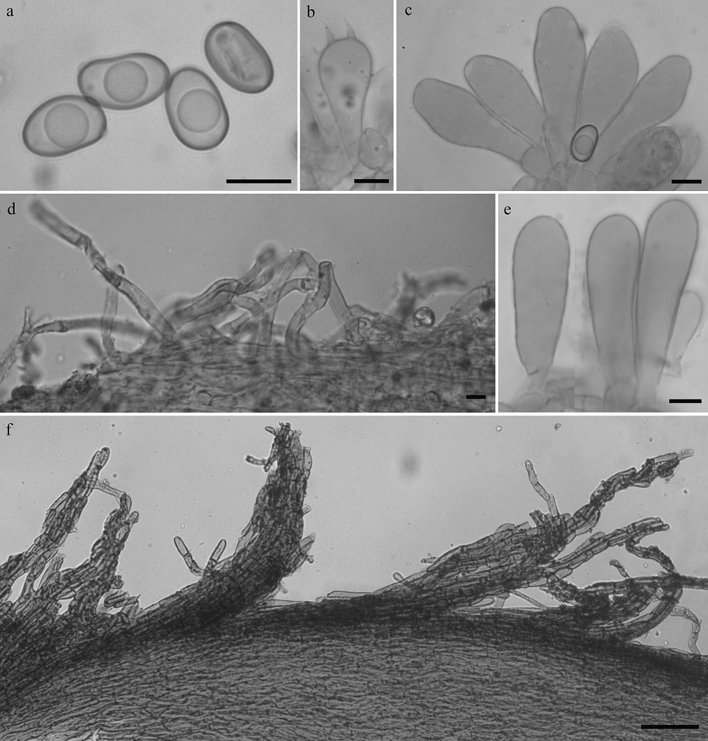
Inocybe brunneosquamulosa (CAL 1308, holotype). a Basidiospores. b Basidium. c Cheilocystidia. d Stipitipellis. e Caulocystidia. f Pileipellis. Scale bars: a-e = 10 μm, f = 100 μm. Photos by K. P. Deepna Latha