Diaporthe cichorii Dissanayake, Camporesi & K.D. Hyde, sp. nov., Index fungorum number: IF553187
Etymology: The specific epithet cichorii is based on the host genus (Cichorium).
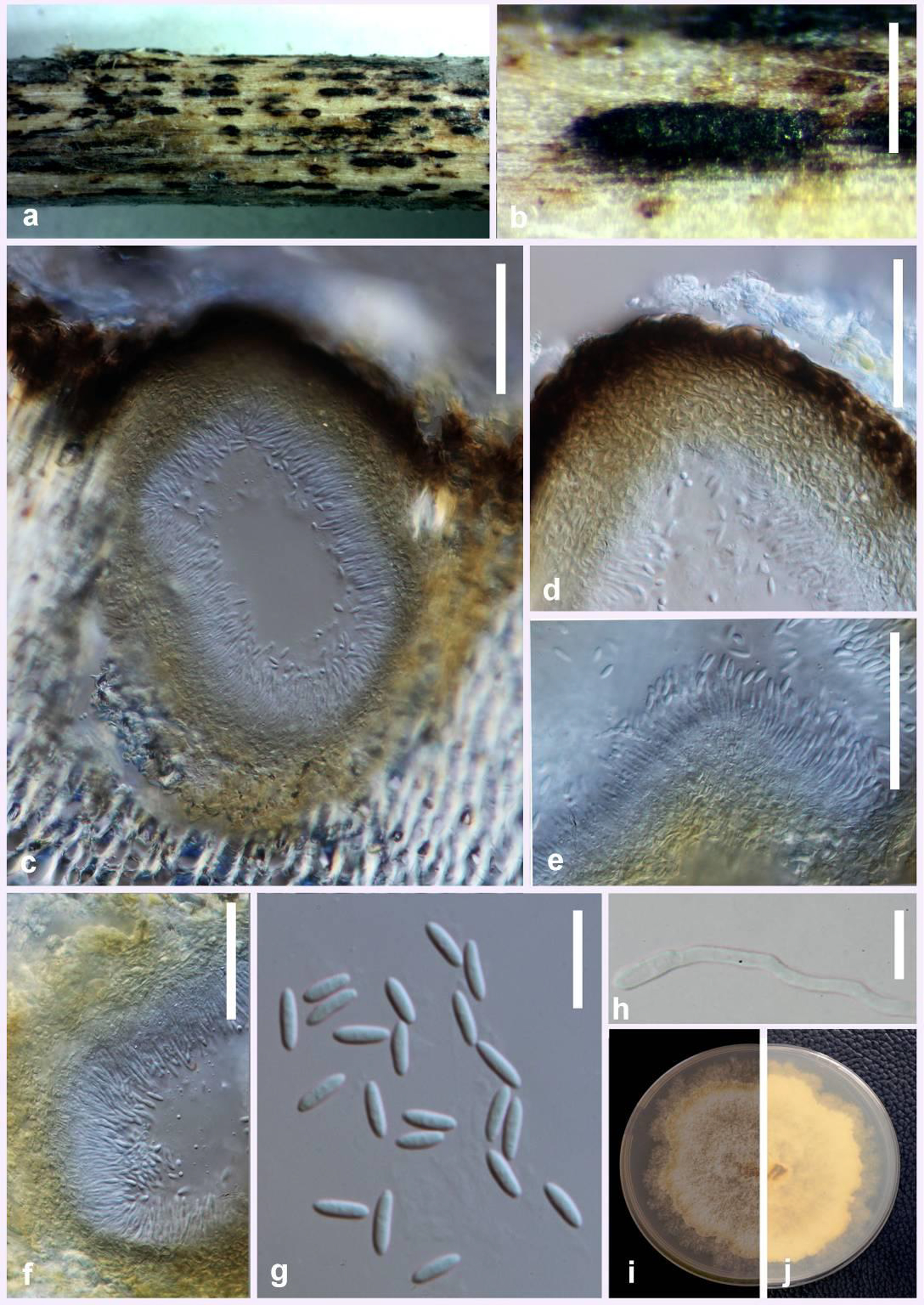
Saprobic on dead aerial stem of Cichorium intybus L. Sexual morph: Not observed. Asexual morph: Conidiomata up to 540 μm in diameter, 390 μm high, superficial, solitary or aggregated, globose to oval, dark brown to black, clustered in groups of 2-5 conidiomata. Peridium 47–58 μm thick, inner layer composed of light brown textura angularis, outer layer composed of dark brown textura angularis. Conidiophores 24–37 × 1.5–3 μm (x̅ = 29 × 3 μm), cylindrical, aseptate, densely aggregated, straight or sinuous, terminal, slightly tapered towards the apex. Conidiogenous cells 7–10 × 2–3 μm, hyaline, subcylindrical, filiform, straight to curved, tapering towards the apex. Alpha conidia 10–14 × 3–4 μm (x̅ = 12 × 3 μm) hyaline, fusiform or oval, both ends obtuse. Beta conidia not observed.
Culture characteristics: Colonies on PDA flat, with an entire edge, mycelium growing in concentric rings, cottony texture, white to smoke-grey; colonies reaching up to 64 mm diameter after one week at 25 °C; reverse buff and isabelline.
Material examined: ITALY, Forlì-Cesena Province, Santa Sofia, on dead aerial stem of Cichorium intybus (Asteraceae), 17 July 2016, Erio Camporesi; (MFLU 16-2168, holotype); extype living culture MFLUCC 17-1023.
Notes: Diaporthe cichorii occurs in a clade separate from D. gulyae, D. cucurbitae and D. subordinaria and differs from D. gulyae by 19 nucleotides in the concatenated alignment, in which 7 were distinct in the ITS region and 12 in the TEF region. Though the sequences of BT region and CAL region are available for D. cichorii, the sequences of those regions are unavailable for D. gulyae. Morphologically, the conidiomata of D. gulyae are up to 3 mm in diam, whereas in D. cichorii they are up to 540 μm in diameter Alpha conidia of D. gulyae are smaller (6.5–9 μm)
compared to those of D. cichorii (10–14 μm).
Fig. Diaporthe cichorii (MFLU 16-2168, holotype). a, b Conidiomata on host surface. c Cross section of conidioma. d Peridium. e, f Conidia attached to conidiogenous cells. g Alpha conidia. h Germinating spore. i, j Culture on PDA after one week. Scale bars: b = 1 mm, c–f = 100 μm, g = 20 μm, h = 10 μm.