Cytospora rosae Senan., Camporesi, & K.D. Hyde, sp. nov., MycoBank: MB821571
Etymology: Named after the host genus Rosa.
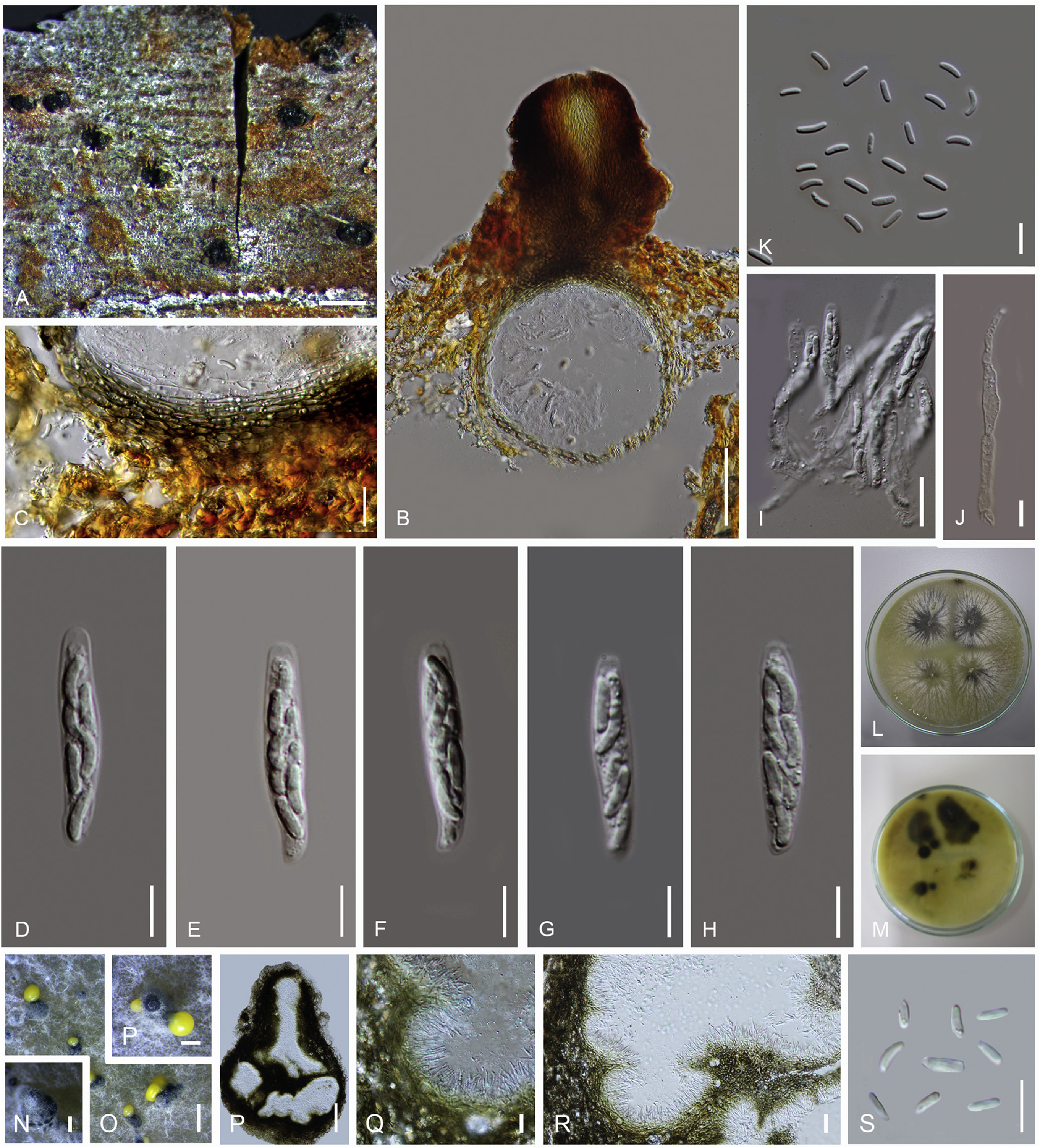
Saprobic on Rosa canina. Sexual morph: Stromata restricted to around the ostiolar neck, black. Ascomata 235–255 μm high, 130–150 μm diam (x̅ = 240 × 140 μm, n = 20), solitary to rarely aggregated, scattered, immersed, globose, brown, coriaceous, ostiolate, papillate. Papilla 127–140 μm high, 70–90 μm diam (x = 135 × 87 μm, n = 20), straight or curved, long, brown, internally covered by hyaline periphyses, wall comprising elongated, thick-walled cells. Peridium 16–23 μm diam (x̅ = 20 μm, n = 20), comprising outer, thick-walled, brown cells of textura angularis and inner, compressed, thick-walled, hyaline cells of textura angularis. Hamathecium comprising septate, hyphae-like, hyaline, 1.5–2.7 μm diam (x̅ = 2.5 μm, n = 20) paraphyses. Asci 20–23 × 3.2–3.7 μm (x̅ = 21 × 3.7 μm, n = 20), unitunicate, 8-spored, clavate, short-pedicellate, apex round, with apical ring. Ascospores 4.2–6.3 × 1–1.5 μm (x̅ = 5.5 × 1.3 μm, n = 20), unito biseriate, unicellular, allantoid, or ellipsoid, hyaline, smoothwalled. Asexual morph: Conidiomata 100–200 μm diam (x̅ = 150 μm, n = 20), solitary to aggregate, immersed, pyriform to subglobose, multi-loculate, black, coriaceous, ostiolate, papillate, peridium folded into centrum. Pycnidial walls 4–7 μm diam. (x̅ = 6 μm, n = 20), comprising small, thick-walled, brown cells of textura angularis. Conidiophores 8–12 × 1.5–2.5 μm
(x̅ = 11 × 2 μm, n = 20), cylindrical, shorter than conidiogenous cells, branched, hyaline. Conidiogenous cells 10–15 × 1–1.5 μm (x̅ = 12 × 1.2 μm, n = 20), phialidic, cylindrical, tapering towards the apices, bearing single conidia at each tip. Conidia 3–5 × 0.5–1 μm (x̅ = 2 × 1 μm, n = 20), aguttulate, elongated to allantoid, slightly curved, aseptate, hyaline.
Culture characteristics: Colonies growing on MEA attained 2 cm within 7 d incubated at 18 °C, filamentous, flat, filiform, middle blackish ash, margin off white, cottony, tiny mycelium clots arrange radially from centre to margin.
Specimen(s) examined: Italy, Province of Forlì-Cesena, Galeata, near Passo delle Forche, on dead branch of Rosa canina (Rosaceae), 15 Apr. 2014, E. Camporesi, IT 1814 (holotype MFLU 17-0885, isotype BBH 42447, cultures extype MFLUCC 14–0845; Province of Forlì-Cesena, Galeata, near Passo delle Forche, on dead branch of Rosa canina (Rosaceae), 4 Jan. 2016, E. Camporesi, IT 1814 (paratype MFLU 15–3596, cultures ex-paratype MFLUCC 17–1664).
Notes: Combined ITS nrDNA, LSU nrDNA, rpb2 and tef1 sequence data in the current study shows that Cytospora rosae forms a distinct clade with high bootstrap support, basal to Cytospora fraxinigena ( Clade 16). Morphologically, Cytospora rosae has unique characters of solitary ascomata and small asci with septate, wide, hyaline, hyphae-like paraphyses.
Fig. Cytospora rosae (MFLU 17–0885). A. Ascomata on substrate. B. Cross section of ascoma. C. Peridium. D–I. Asci. J. Paraphysis. K. Ascospores. L. Colony on MEA upper surface. M. Colony on MEA lower surface. N–P. Conidiomata on MEA. P. Horizontal cross section of conidioma. Q–R. Peridium with conidiogeneous cells, conidiophores and conidia. S. Conidia. Scale bars: A, P–N = 500 mm, B = 50 mm, C, I, Q–R = 10 mm, D–H, J, K, S = 5 mm, P =100 mm, O = 1 mm.