Coronophora myricoides H.X. Wu & K.D. Hyde, sp. nov., Index Fungorum number: IF552180
Etymology: Referring to the resemblance of the ascomata (under the stereomicroscope) to the berries of Myrica
rubra (waxberry).
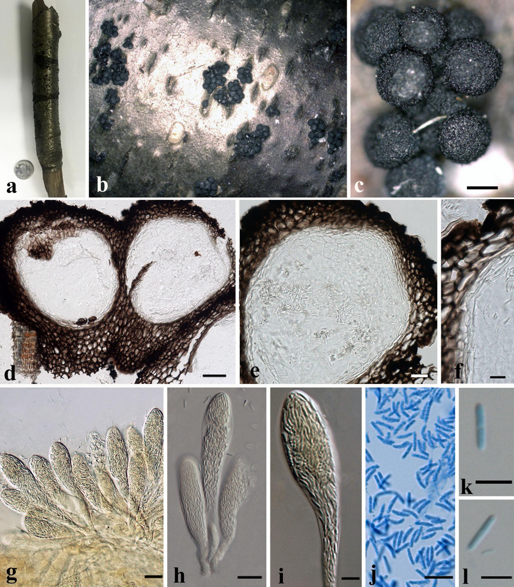
Saprobic on the surface of dead wood. Asexual morph Undetermined. Sexual morph Ascomata 373–395 μm high × 282–340 μm diam. (x = 383 × 308 μm, n = 10), perithecial, superficial or erumpent through bark of host, clustered in small to large groups, subglobose, surface tuberculate, dark brown to black, lacking ostioles. Peridium 27–135 μm (27–56 μm at the apex, 93–135 μm at the base), composed of five layers of dark brown-walled cells of textura globosa, inner layer of hyaline pseudoparenchymatic cells, slightly melanized on the outer surface. Paraphyses not seen. Asci 102–147 × 11–27 μm (x = 119 × 22 μm, n = 15), numerous, polysporous, unitunicate, cylindrical to clavate, pedicel 11–38 × 4–7 μm. Ascospores 6–8 × 1–2 μm (x = 7 × 1.7 μm, n = 30), numerous, hyaline, 1-celled, cylindrical or ellipsoidal, narrowly rounded at both ends, smooth-walled.
Material examined: CHINA, Yunnan Province, Jing Dong, on dead wood of unidentified plant, 13 September 2013, Wu Hai Xia (IFRD 9201, holotype).
Notes: The genus Coronophora is typified by Coronophora gregaria and there are 28 epithets in Index Fungorum (2016). Kirk et al. (2008) estimated that there are five species in Coronophora from wood. There are two sequences for Coronophora gregaria in GenBank. In this paper, we introduce a new species of Coronophora; C. myricoides which differs from C. gregaria in the shape of the ascomata and also the shape of ascospores. The ascospores of Coronophora myricoides (6–8 × 1–2 µm) are smaller the type species (8–12 × 2–3 µm). We directly extracted DNA using ascomata and sequencing. We add the molecular data to better understand the generic and familial circumscriptions.
Fig. Coronophora myricoides (holotype). a Herbarium material. b, c Appearance of ascomata on wood. d, e Vertical sections of ascomata. f Peridium. g–i Asci. j–l Ascospores. Scale bars c = 200 µm, d = 50 µm, e, g, h = 20 µm, f, i–l = 10 µm.