.
Biscogniauxia marginata (Fr.) Pouzar, Česká Mykol. 33(4): 216 (1979)
Index Fungorum Number: IF 309561; Facesoffungi number: FoF 000299
Holotype – UPS: BOT: F-175466
Saprobic on wood. Sexual morph Ascostromata 3.5–7×2–3 mm(x=5.6×2.8 mm), raised–discoid, globose, with concave surface, distinct raised margins, outer dehiscing layer, surface black, woody layer immediately beneath the stromatal surface and between perithecia with carbonaceous tissue encasing each ostiole, tissue beneath perithecia comprising with host tissue, carbonaceous, perithecia obovoid, 0.2–0.5×0.5–1 mm (x=0.4×0.8 mm), thick-walled, light brown inner cell layers, outer cell layers carbonaceous, ostioles slightly umbilicate with punctuate openings, with white residues. paraphyses numerous, between asci, filamentous, septate, 3–5×50μm or longer (x=4.4×50μm). Asci (145–)160–200(–204)×(8.3)9–12(12.2) μm (x=176×13μm, n=20), 8-spored, unitunicate, cylindrical, pedicellate, with apical ring bluing in Melzer’s reagent, discoid, 0.5–0.7×1.5–2μm (x=0.6×1.7μm, n–20). Ascospores (12.4–)13–16.5(–16.8)×10–13.5μm (x=15×11.7μm, n=30 ), uniseriate, one-celled, globose to subglobose, with broadly rounded ends, dark brown to black, with sigmoid germslit the entire spore-length, smooth-walled. Asexual morph Sporulating regions scattered over entire central part of the colony, brown-vinaceous (84) after 4 weeks. Conidiogenous structure nodulisporium-like, arising as roughened masses of hyphae, hyaline. Conidiogenous cells 50–60×4–5μm (x=56×4.4μm), hyaline, finely roughened. Conidia 6–8×3.5–4μm (x=7.2×3.8μm), hyaline, smooth to finely roughened, ellipsoid.
Culture characters – Colonies on OA at 25–28 °C reaching the edge of 6 cm Petri-dish in 14 days, whitish, velvety to felty, azonate, with diffuse margins, reverse at first straw (46), later developing into dark brown colonies after 3–4 weeks.
Material examined – FRANCE, on wood, 16 April 2012, Erio Gardiennet AXL 001 (MFLU 13–0099), living cultures, MFLUCC 12–0740.
GenBank Accession Numbers – ITS: KJ958407; LSU:KJ958408; RPB2: KJ958409.
Notes – Biscogniauxia marginata and B. baileyi are similar in their stromatal characters. Both have subglobose ascospores which make them different from many of the other species in the genus. However, B. marginata has globose ascospores with a sigmoidal germ slit, while B. baileyi has straight germ slit. Biscogniauxia marginata has a nodulosporium-like asexual morph as observed here. Callan and Rogers (1986), Petrini and Müller (1986) and Ju and Rogers (1996) mentioned the asexual morph as nodulosporium–like, while Whalley and Edwards (1985) recognised the asexual morph as geniculosporium-like. In GenBank there is only one ITS sequence of belonging to B. marginata and we add ITS, LSU, RPB2 and β-tubulin gene data from an authentic specimen.
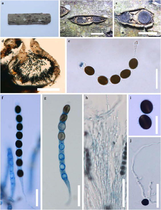
Fig. 1 Biscogniauxia marginata (MFLU 13–0099). a Habit b, c Concave stromatal surface in with carbonaceous tissue d Cross section of stromata showing the perithecium and ostiole e J+ apical apparatus in Melzer’s reagent f Mature ascus in Lactophenol Cotton Blue g Ascus in Lactophenol Cotton Blue showing sigmoid germ slit h. Paraphyses in water i Ascospores in water j Germinating ascospore. Scale bars: b, c=5 mm, d=0.2 mm, e–h=20μm, i, j=15μm.
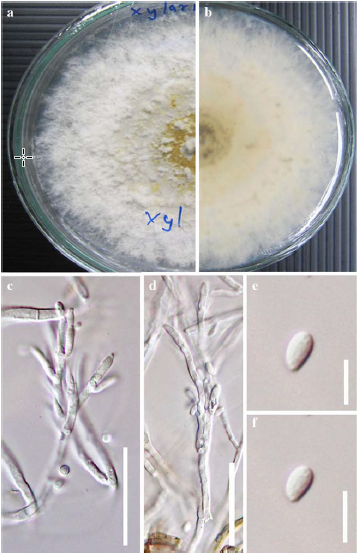
Fig. 2 Biscogniauxia marginata in MEA after 2 weeks a From above – white colony b From below – straw (46) c Conidiogenous structure d Conidiogenous cells e, f Conidia. Scale bars: c, d=30μm, e, f=5μm.