Amanita atrobrunnea Thongbai, Raspé & K.D. Hyde
Index Fungorum number: IF 551652 Facesoffungi number: FoF 02070
Etymology: the epithet refers to the dark brown colour of the pileus
Holotype: MFLU 15 –1415 Pileus 120 mm in diam., conic to paraboloid when young, then plano-convex, becoming convex and broadly umbonate when mature, dark brown to chestnut brown (6 F7, 6 F8), darker in the center, paler and becoming teakbrown to leather brown (6F5) towards the margin, minutely rimose, sub-viscid when wet; margin lacking striations, slightly appendiculate, sometimes with scattered annulus remnants; context 1 mm thick at mid-radius, white. Lamellae free, white, crowded, up to 8 mm high; lamellulae attenuate,with two to three series. Stipe 170×15 mm, slender, slightly tapering upwards, white to pale yellowish, finely fibrillo-squamulose; context white, solid, unchanging when bruised. Bulb 15–25 mm wide,inconspicuous, subfusiform, white (1A1). Volva limbate, slightly firm, up to 20 mm high, white (1A1). Annulus membranous, easily broken, white. Odour absent. Lamellar trama bilateral; mediostratum 30–35μm wide, composed of ellipsoid to fusiform, 35–45×10–18μm cells, mixed with abundant, filamentous 3–6 μm wide, branching hyphae. Subhymenium 20–35μm thick, with two to three layers of subglobose to irregularly-shaped cells, 12–25×10– 15μm. Basidia 36–41×9–12μm, 4-spored, clavate, thinwalled; sterigmata 4–6μm long. Basidiospores 7.3–8.3– 9.5×5.4–6.6–7.8μm, Q=1.15–1.26–1.46 (N=40), broadly ellipsoid to ellipsoid, thin-walled, colourless, amyloid, smooth, with small apiculus. Lamellaredge composed of numerous, subglobose, (15–25×8–18μm) cells, and rare filamentous, thin-walled, hyaline, 3–9μm wide hyphae. Pileipellis 90–100μm thick, composed of two distinct layers, the upper laye gelatinized, made up of radially arranged, thinwalled, filamentous, 3–8 μm wide, colourless hyphae, with inflated, sometimes cylindrical, rarely subglobose to elliptical terminal cells; the lower layer mostly non-gelatinized, composed of filamentous, sometimes branching, 4–10μm wide hyphae with palebrown pigment, mixed with abundant inflated cells. Velar remnants from stipe base composed of thinwalled to slightly thick-walled, filamentous, 3–8μm wide hyphae, mixed with abundant inflated cells, with yellowish to pale brown intracellular pigments. Annulus composed of thin toslightlythick-walled, filamentous, 3–8 μm wide, branching hyphae, mixed with ellipsoid to subglobose, hyaline, inflated, thin-walled cells. No clamps observed in any tissue.
Habitat: Terrestrial in forest dominated by Fagaceae species.
Material examined: THAILAND, Chiang Mai Province, Doi Saket District, Sub-District Tepsadet, N18° 57′ 1.0016″ E99° 20′ 1.0452″, 30 June 2014, collector B. Chuankid, BZ– 2014–09 (MFLU 15–1415, holotype)
Notes: Amanita atrobrunnea is a member of Amanita subgenus Lepidella (J.-E. Gilbert) Veselý, section Lepidella (Bas 1969). Remarkable features of A. atrobrunnea are the dark brown pileus, the broad umbo at the disc, the slender basidiocarp, the absence of membranous velar remnants on the pileus, even when young, the abundant inflated cells in the pileal surface and the broadly elipsoid to ellipsoid basidiospores. The most morphologically similar species are A. manginiana sensu W.F. Chiu and A. pseudoporphyria Hongo, which share several characters with A. atrobrunnea, such as an inconspicuous bulb, dark pileus, and velar remnants on the pileus consisting of inflated cells (Zhang et al. 2010). However, A. atrobrunnea can easily be distinguished from A. manginiana and A. pseudoporphyria by its distinctive umbonate pileus at maturity. In addition, the inflated cells of the pileipellis, a key character of A. atrobrunnea, are not present in the other species. Like A. atrobrunnea, A. pallidorosea P. Zhang & Zhu L. Yang possesses a conspicuous umbo, but the pallid rose colour of latter is very different. Amanita manginiana and A. pseudoporphyria were initially placed in section Phalloideae (Hongo 1982; Yang 1997; Zhang et al. 2004, 2010). However, recent phylogenetic analyses clearly showed that both species belong to section Lepidella (Cai et al. 2014). Our molecular phylogenetic analysis indicates that A. atrobrunnea is a sister species to A. manginiana and A. pseudoporphyria.
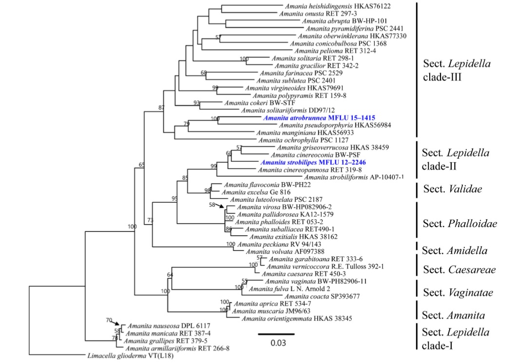
Phylogram inferred by Maximum Likelihood analysis of LSU sequences. Bootstrap support values greater than 50 % are indicated above the nodes. New taxa are in blue and species for which obtained sequences are based on type material have names in bold. The tree is rooted with Limacella glioderma
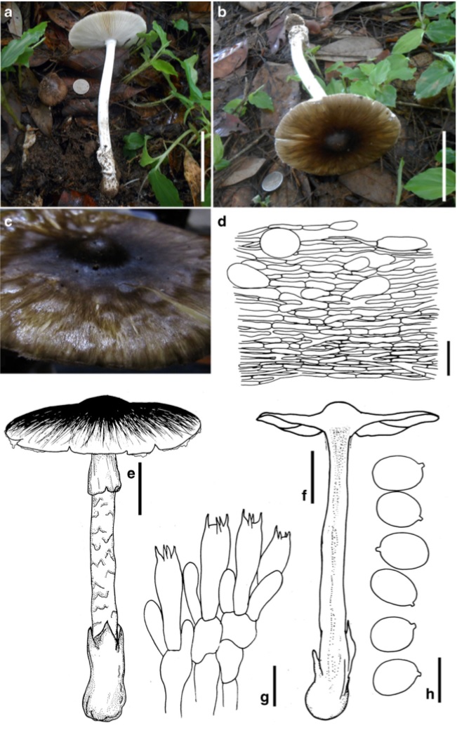
Amanita atrobrunnea (holotype) a–c Basidiome d Radial section of pileipellis e, f Basidiome g Basidia and subhymenium h Basidiospores. Scale bars: a, b=8 cm, d=20μm, e, f=30 mm, g=20μm, h=10μm