Lopadostoma fagi Jaklitsch, J. Fourn. & Voglmayr, in Jaklitsch et al. Persoonia, Mol. Phyl. Evol. Fungi 32: 63 (2014)
Index Fungorum number: IF 803805; Facesoffungi number: FoF 01897
Saprobic on dead twigs and branches of Fagus sylvatica L. Sexual morph: Stromata 700–1500 × 300–800 µm (x̅ = 1200 × 500 µm, n = 30), effused–pulvinate, immersed in the host and erumpent from bark, bluntly conical, surrounded by a narrow, black, carbonized encasement, appearing as a black line, ectostromatic disc visible as a clypeus and surrounded by reddish brown bark surface, convex, raised, dark grey, entostroma dark, usually black, multi-peritheciate. Ostiole papillate with inconspicuous ostiolar openings. Perithecia 200–800 × 150–680 µm (x̅ = 400 × 520 µm, n = 30), 2–5 clustered in valsoid groups, mono-distichous, subglobose to flask-shaped, at the periphery inclined toward the center, tissue between perithecia loosely arranged, composed of white fungal tissue, mixed with light coloured bark cells, tissue beneath the perithecia compact, black, with short convergent ostiolar necks. Paraphyses are numerous, long, apically free, 1–3.5 µm wide (x̅ = 3 µm, n = 30), rarely branched. Asci 79.8–103.5 × 4.6–6.1 µm (x̅ = 91 × 5.3 µm, n = 30), unitunicate, 8-spored, cylindrical, pedicellate, ellipsoidal-discoid, inconspicuous apical apparatus typically 2–3.5 µm wide. Ascospores 7.5–8.7 × 3.2–4.2 µm (x̅ = 7.9 × 3.9 µm, n = 30), uniseriate, oblong to narrowly ellipsoid, symmetrical to slightly inequilateral, unicellular, lacking a dwarf cell, at first hyaline, turning pale brown and dark brown at maturity, smooth-walled, with a straight germ slit across the entire spore length present, when immature with 2 large guttules.
Culture characteristics – Colonies on Difco OA very slow growing, reaching the edge of 9 mm Petri-dishes in 3–3 ½ months at 25–27 °C, at first whitish, velvety, azonate, with slightly lobed margins, developing sporulating regions as light-yellow mycelial masses in culture; later, reverse turning light orange.
Specimen examined – Italy, Province of Forlì-Cesena, Campigna – Santa Sofia, dead branch of Fagus sylvatica L (Fagaceae), 19 September 2014, E. Camporesi, (MFLU 15-2600, KUN), living culture (MFLUCC 15-0008, KIBCC).
Notes – Lopadostoma fagi is reported as a common species in corticated branches of Fagus sylvatica from Europe (Jaklitsch et al. 2014). This specimen was collected from Italy on the same host. According to the morphological description of L. fagi provided in Jaklitsch et al. (2014) it is characterized in having distinctly papillate ostioles and smaller, narrower ascospores with a straight, full length germ slit. Similar morphological traits were observed in our collection. However, we observed 2–5 perithecia, in stroma more or less in a valsoid configuration and rarely distichous, even though according to Jaklitsch et al. (2014) it is mentioned as (3–) 6–8 in a cluster and monostichous. In addition there are slight variations in the measurements of perithecia, asci and ascospores which can be due to the geographical variations and environmental conditions. The slow germination of ascospores (3 weeks on MEA and 4–5 weeks on OA) later resulted in whitish colonies is similar to those observed by Jaklitsch et al. (2014) who mentioned as 1–4 weeks for initial germination of ascospores on MEA. Due to these morphological similarities between our collection and the previously studied specimens, we have identified this species as L. fagi, which is also confirmed by the molecular studies.
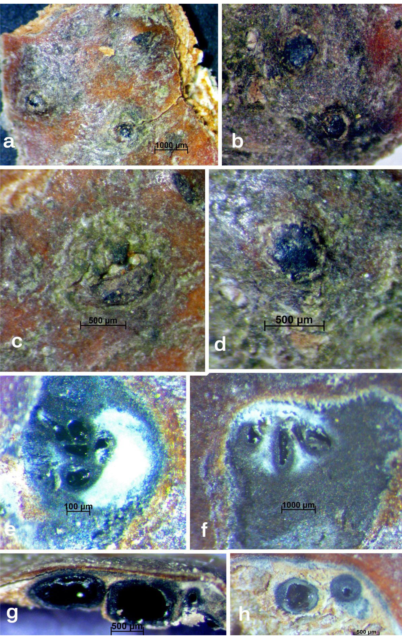
Fig. 1 Lopadostoma fagi a-c. Surface of stromata on host, d. ectostromatic disc, e, h. transverse sections of stromata, f, g. vertical sections of stromata. Scale bars: a = 1000 µm, b–d = 500 µm, e = 100 µm, f = 1000 µm, g, h = 500 µm.
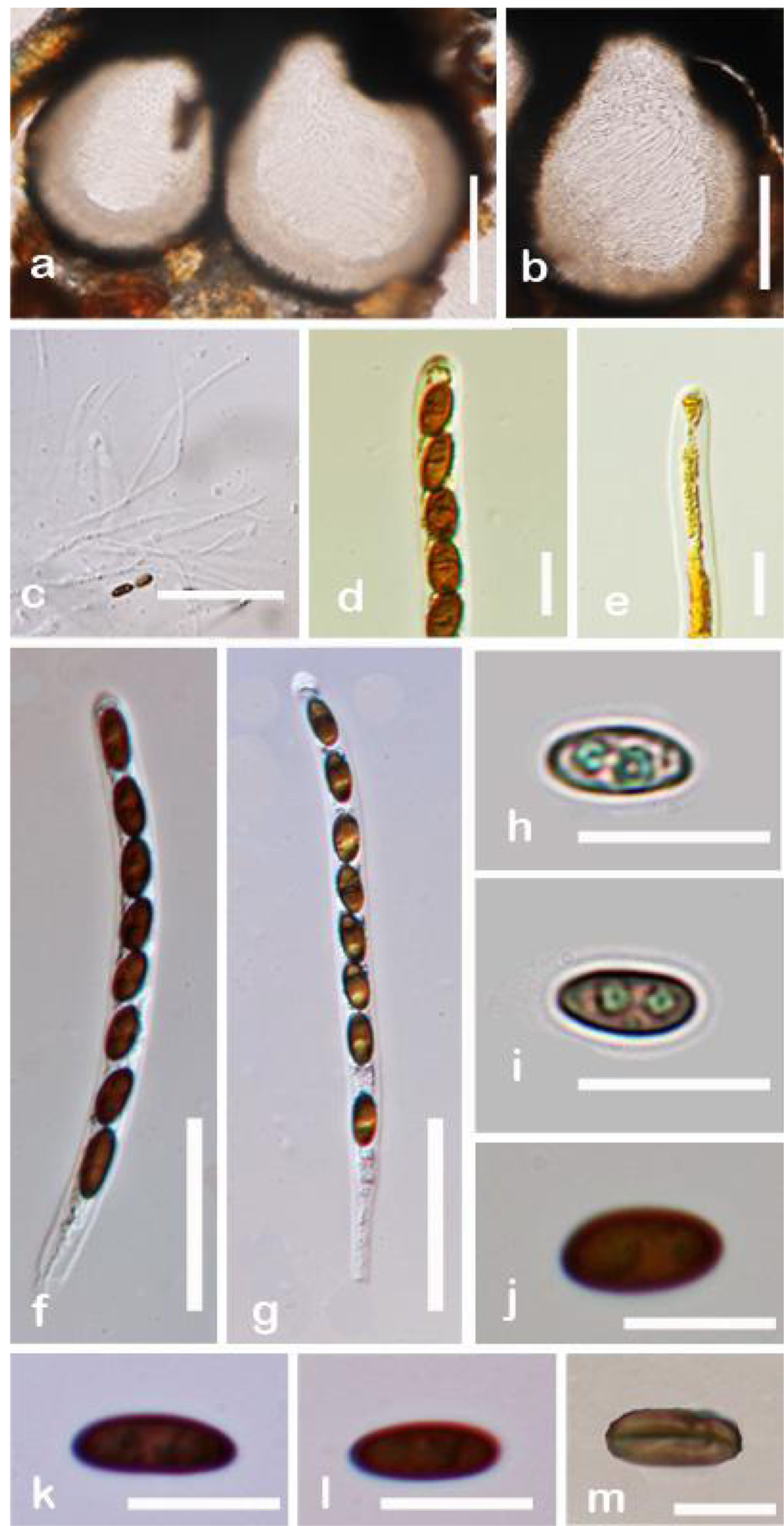
Fig. 2 Lopadostoma fagi a, b. Vertical sections of stromata, c. Paraphyses, d, e. Asci with inconspicuous apical apparatus (d-in Melzer’s reagent, e-in Lugol solution), f, g. Mature asci, h-i. Immature ascospores, j-l. Mature ascospores, m. Ascospore with straight germ slit. Scale bars: a, b = 300 µm, f = 30 µm, d-e = 10 µm, f, g = 30 µm, h-m = 10 µm.