Graphostroma platystomum (Schwein.) Piroz., Can. J. Bot. 52(10): 2131 (1974)
Index Fungorum Numbers: IF 626523; Facesoffungi Number: FoF 02969
Basionym: Sphaeria platystoma Schwein.
Saprobic on trunks, branches, and twigs of several genera of deciduous trees. Sexual morph: Ectostroma young stromata cinnamon colour, hard, brittle, carbonaceous, composed of dark hyphae with periderm cells, at maturity ectostromatic crust separates, exposing the entostromatic ‘‘disc’’, ostiolate, dark cinnamon to chestnut brown, pulvinate, with a thin, sterile, fimbriate margin. Ascomata immersed, compressed globose, bottle-shaped, with long necks, blackish brown, more rarely pyriform, or obpyriform, usually monostichous. Peridium composed of indistinct, hyaline, tangentially flattened cells. Paraphyses hyaline, very thin-walled, inconspicuous, narrowly ventricose, 3-septate, tapering gradually to a pointed apex. Asci 8-spored, unitunicate, clavate, subsessile to short-pedicellate, apex rounded to subtruncate, with a minute J+ apical ring, inconspicuously bluing in Melzer’s reagent, discoid. Ascospores overlapping biseriate, unicellular, suballantoid, tapered at both ends, inequilateral, hyaline, thin-walled, without germ slit. Asexual morph: Nodulisporium-like. Conidiogenous cells thin-walled, roughened at the base, apex swollen into a clavate or globose vesicle, dilute straw coloured towards the base, hyaline above, bearing up to 12 small, inconspicuous denticles, arising singly from the apex or sides of more or less erect mycelial hyphae, or in whorls of three to five from apices of erect hyphae. Conidia pyriform to turbinate, rounded above, hyaline, unicellular, strongly attenuated towards a minute thickened and refractive basal scar, containing a single, large guttule.
Material examined – FRANCE, Ariege, Riverenert, Les Cravives, 700 m, on bark of Quercus sp. 25 Jul. 2011, J. Fournier (JF 11090).
Notes – Pirozynski (1974) mentioned that he was not aware of the existence of the original collection or any other subsequent collections. He observed and listed several specimens related to Graphostroma platystomum, even though none of them have been typified. Most of these specimens are deposited in DAOM with a few at K. We have borrowed and observed two specimens observed by Pirozynski (1974). One specimen (DAOM 36086) contains little material and was in poor condition. We observed only a few dried ascospores which are hyaline and somewhat allantoid with tapered ends matching the original description by Pirozynski (1974). The other specimen (DAOM 36091) contains a different fungus with bitunicate asci. Therefore, we have provided a complete illustration from authentic material and compared with the illustration redrawn from Pirozynski (1974).
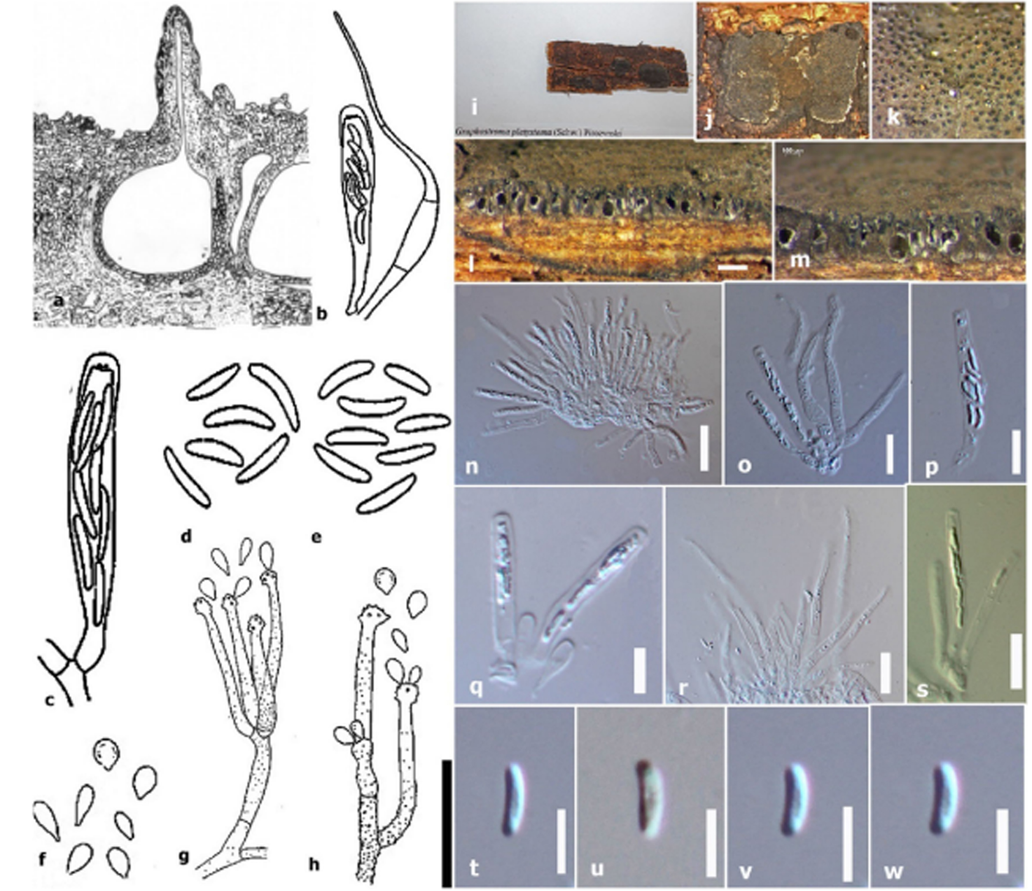
Fig 1. Graphostroma platystomum (a–h redrawn from Pirozynski 1974). a Ascoma in cross section. b Ascus from DAOM 75729. c Ascus and paraphyses from DAOM 127673. d Ascospores from DAOM 36086. e Ascospores from DAOM 65174. f Conidia. g Conidiophores and conidia from DAOM 36091. h Conidiophores and conidia from DAOM134022. Scale bars: b–f = 20 μm; (i–w JF 11090) i Stromal habitat. j Stromal surface. k Stromal surface showing ostioles. l, m Cross section of stroma showing ascomata encased in stromal tissues. n–q Asci. r Paraphyses. s Apex of asci in Melzer’s reagent. t–w Ascospores. Scale bars j = 500 μm, k, l = 200 μm, m = 100 μm, n–s = 20 μm, t–w = 10 μm.