Colospora andalasii Miettinen & Spirin
MycoBank number: MB 813994 Facesoffungi number: FoF00995
Etymology: After Andalas University, the leading botanical research institution in Sumatra.
Holotype: Miettinen 13096
Basidiocarps annual, resupinate, leathery, up to 15 cm in widest dimension, up to 0.3 mm thick. Margin compact, sterile. Hymenial surface pale cream coloured, odontoid, in older parts with vinaceous stains; spines (sterile hyphal pegs) solitary, regularly arranged, up to 200–300×40–60 μm, 5–7 per mm, with sharp apices. Subiculum (thickness excluding the spines) 150–250 μm. Hyphal structure dimitic (amphimitic), generative hyphae clamped. Skeletal hyphae dominating in all parts of basidiocarps, irregularly arranged in subiculum, (1.8)2–2.8(3.4) μm( n=34/1), subparallel in spine trama, (2)2.6–2.9(3.2)μm(n=20/1), branched and tapering but sparingly, thick-walled, with a capillary, rather indistinct lumen (one sixth of hyphal diam. or less), acyanophilous, faintly yellowish or hyaline in Melzer’s reagent.Generative hyphae thin-walled, 1.8–2.8 μm. Coarse crystals abundant in subiculum, up to 20 μm in widest dimension, mostly of square or rhomboidal shape; also fine sandlike encrustation. Hymenium covers spines only close to their base and does not extend close to the tip. Dendrohyphidia present in hymenium, usually not projecting, 2.5–4 μm in diam., with short blunt branches, collapsing easily. Basidia utriform, 4-spored, 30–40×9–10 μm, with oil drops inside, sterigmata subulate, up to 12×2 μm. Basidiospores slightly thickwalled, finely ornamented (covered by minute warts), biapiculate, apical parts distinctly tapering and refractive, with numerous oil drops inside, faintly cyanophilous, showing small amyloid patches in apices, 14.7–18.8×(5.7)6–7.3(7.8), L=16.99 μm, W=6.65 μm, Q=2.56 (n=32/1).
Material examined: Colospora andalasii: INDONESIA, Sumatera Barat, Padang, Limau Manis, dry fallen angiosperm branch, 15 July 2008 Miettinen 13096 (ANDA holotype, H), INSD KT361629. Epithele alba: BRAZIL, São Paulo, Campinas, Moji-Guaçu, 29 January 1987 Ryvarden 24517 (H ex O). Epithele subfusispora: BRAZIL, São Paulo, Campinas, Moji-Guaçu, 23 January 1987 Ryvarden 24554 (H ex O). Epithele typhae: ESTONIA, Valgamaa, Otepää, Valkjärva, Carex acutiformis, 10 September 2012 Kotiranta 25248 (H). Erythromyces crocicreas; INDONESIA, Sumatera Barat, Padang, Limau Manis, dicot log, 16 July 2008 Miettinen 13157 (ANDA, H), INSD KT361630. Skeletohydnum nikau: NEW ZEALAND, Auckland, Waitakere Ranges, Rhopalostylis sapida (fallen twig), 24 February 1984 Buchanan 84/016 (H ex O). Theleporus venezuelicus: VENEZUELA, Estado Bolivar, Sifontes, Tumeremo, 17 November 1994 Ryvarden 35205 (O, holotype), INSD KT361631.
Notes: Colospora andalasii is very similar morphologically to Epithele citrispora described from Gabon. The main separating character is the ornamentation of spores–clearly visible in C. andalasii, but not reported for E. citrispora. Boidin and Gilles (1998) produced an ITS sequence of a paratype of E. citripora. Even if we do not have control over the quality of the E. citrispora sequence, the ITS difference between the two seems convincingly large(>10%) to exclude conspecificity. A few Epithele species have ornamented spores: E. alba (Viegas) Boidin et al.,E. interrupta Bres. and E.subfusispora (Burds. & Nakasone) Hjortstam & Ryvarden (Hjortstam and Ryvarden 2005). Their hyphal structure is different from that one of Colospora, considered as monomitic (Hjortstam and Ryvarden 2005) or dimitic with ‘microbinding hyphae’ (Nakasone 2013).
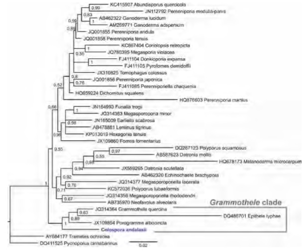
Position of Colospora within the core polyporoid clade (Polyporales, Basidiomycota). Consensus phylogram of the 4503 trees retained in the Bayesian analysis of nrDNA ITS and LSU. Numbers represent Bayesian posterior probabilities.
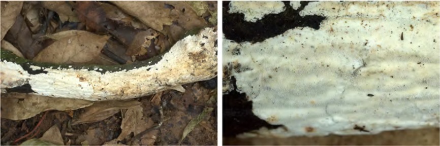
Photographs of Colospora andalasii (holotype) in the field.
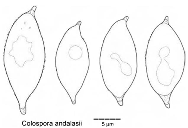
Basidiospores of Colospora andalasii (isotype) drawn in Cotton Blue.