Anthostomella forlicesenica Daranagama, E. Camporesi & K.D. Hyde, in Daranagama, Camporesi, Tian, Liu, Chamyuang, Stadler & Hyde, Fungal Diversity 73: 214 (2015)
Index Fungorum number: IF 811101; Facesoffungi number: FoF 00321
Etymology – In reference to the name of the province (Province of Forlì-Cesena) where the material was collected.
Holotype – MFLU 14-0227
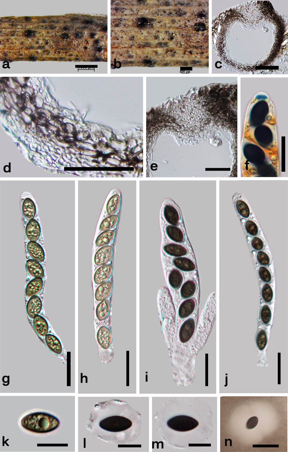
Saprobic on stems, branches and twigs. Sexual morph: Ascomata immersed, visible as conical blackened dots, dark brown-black, mostly solitary, in cross-section 200–250 µm diam. and 250–280 µm high (x̄ = 237.5 × 265 µm, n = 10), globose, with a central periphysate ostiolar canal, 30–40 µm diam. 40–50 µm high (x̄ = 37.3 × 45.7 µm, n = 10). Peridium 15–20 µm diam. (x̄ = 18.5 µm, n = 10) with several cell layers, outwardly comprising dark brown cells of textura globulosa and inwardly comprising hyaline cells of textura angularis. Paraphyses 5 µm diam. (x̄ = 5 µm, n = 30), filamentous, septate, few. Asci 95–120 × 13–16 µm (x̄ = 117.5 × 15 µm, n = 30), 8-spored, unitunicate, cylindrical, short-pedicellate, apically rounded, J+ apical apparatus, discoid, 2–2.5 × 0.5–1 µm (x̄ = 2.3 × 0.8 µm, n = 30). Ascospores 13–15 × 6–7 µm (x̄ = 14.5 × 6.5 µm, n = 40), uniseriate or overlapping uniseriate, brown, equilateral with parallel sides, unicellular, lacking a germ slit, with a 6.5–7.5 µm (x̄ = 6.8 µm, n = 40) diam. conspicuous mucilaginous sheath. Asexual morph: Undetermined.
Culture characteristics – Colonies on Difco OA at 25–27 oC reaching 5 cm in 4 weeks, at first whitish, felty, azonate, with diffuse margins, reverse turning citrine (13) or yellow.
Specimens examined – ITALY, Province of Forlì-Cesena, Monte Riccio – Bagno di Romagna, on Spartium junceum L. (Fabaceae), 6 January 2013, E. Camporesi IT995 (MFLU 14-0227, holotype), ex–type cultures, MFLUCC 14-0007; ibid, 6 January 2013, E. Camporesi IT995 (PDD, isotype). Province of Forlì-Cesena, Corniolo, on Spartium junceum L. (Fabaceae), 5 February 2013, E. Camporesi IT995–bis (MFLU 14-0228, PDD, paratypes), ex–paratype culture, MFLUCC 14–0558.
Notes – Anthostomella forlicesenica and A. zongluensis K.D. Hyde are similar. However, A. zongluensis can be distinguished from A. forlicesenica as the former has minute globose ascomata usually not visible to the unaided eye and ascospores that are ellipsoidal, verruculose and grey-brown and have parallel sides (Hyde 1996). In A. forlicesenica ascomata are visible as conical areas and in cross section are subglobose. Ascospores of A. forlicesenica are ellipsoidal, dark brown or black with pointed ends, have a large mucilaginous sheath and lack verruculose walls.
Fig. 1 Anthostomella forlicesenica (MFLU 14–0227, holotype). a Habit on wood. b Ascomata in wood. c Cross section of an ascoma. d Peridium. e Clypeus. f Asci in Melzer’s reagent with J+, discoid apical apparatus. g, h Immature asci in water. i, j Mature asci in water k Immature ascospore. l, m Mature ascospore in water. n Ascospore in Indian ink showing the mucilaginous sheath. Scale bars: a = 1 mm b = 500 μm, c, d, e = 50 μm, f–n = 10 μm