Mimicoscypha T. Kosonen, Huhtinen & K. Hansen, gen. nov. Fig. 14f – h, 15 –18
MycoBank number: MB 835735; Index Fungorum number: IF 835735; Facesoffungi number: FoF 14070;
Etymology: For mimicking two other genera, Eupezizella and Resino- scypha.
Type species: Mimicoscypha lacrimiformis (Hosoya) T. Kosonen, Huhtinen & K. Hansen.
Included species: M. lacrimiformis, M. mimica, M. paludosa.
A genus with light-coloured, hairy apothecia on wood, bryophytes and plant debris. Hairs with 1– 3 septate, thin- to somewhat thick-walled, hyaline, mostly smooth, sometimes with hyaline to yellowish, superficial resin; inside the hairs hyaline, refractive globules variably present in living, occasionally in dried material, amyloid nodules not present. Ectal excipulum of textura angularis – prismatica, typically without dextrinoid reactions, but may show strongly amyloid nodules inside the cells. Asci arising from croziers or simple septa, apical pore MLZ+, 8-spored. Ascospores ellipsoid to oblong-ellipsoid, aseptate to 1-septate. Paraphyses cylindrical.
Notes: — Mimicoscypha earns its name by sharing morphological features with both Eupezizella and Resinoscypha, despite it is clearly distinct from these genera based on our multi-gene data (Fig. 2). Delimitation may prove difficult if only morphological features are used. It differs from Eupezizella in the septate hairs, and from both Eupezizella and Resinoscypha in not showing amyloid nodules in the hairs (see further in Notes under Resinoscypha below). These blunt-haired, hyaloscyphaceous taxa have been vaguely placed for decades, and a strongly supported placement has not been offered until now. Our solution is based on a combination of morphological characters and molecular phylogenetic results. Mimicoscypha is closely related to the genus Olla and Hyalopeziza nectrioidea (Fig. 2) although species of Olla and H. nectrioidea have very different excipular hairs (see Fig. 14c – d, f). The long slender, thin- to somewhat thick-walled, hyaline hairs, without glassy elements (typical for Olla) or a dextrinoid reaction (typical for both Olla and H. nectrioidea), support the creation of this new genus.
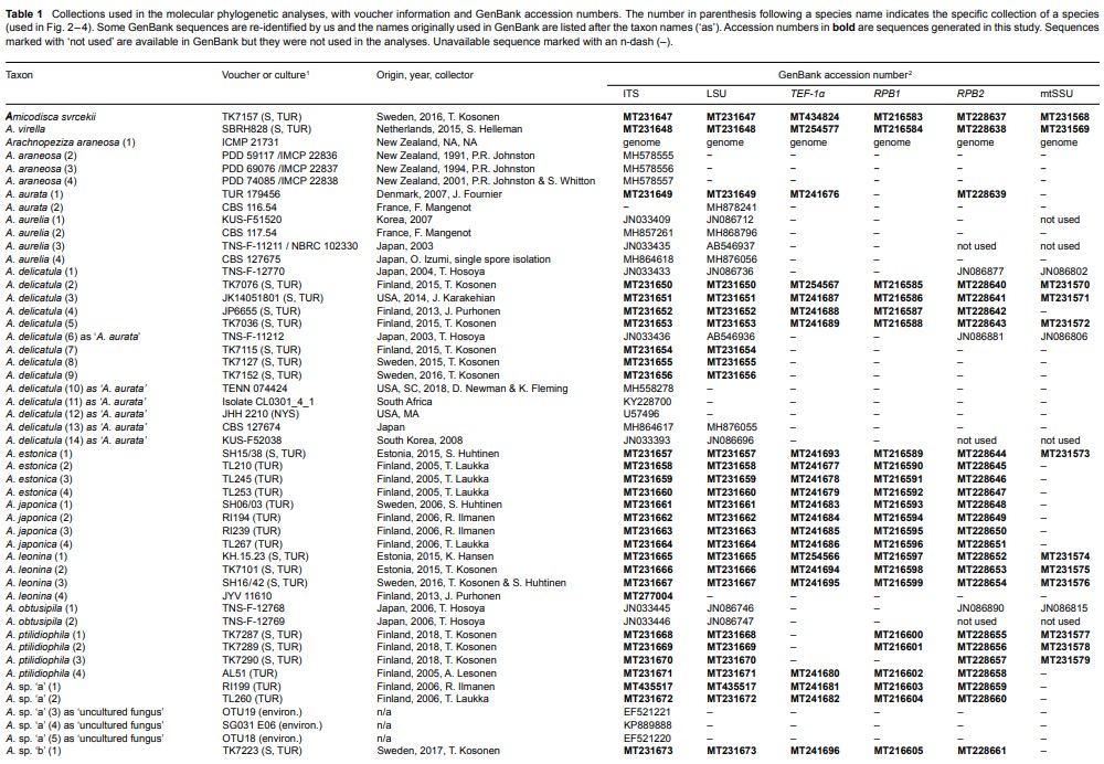
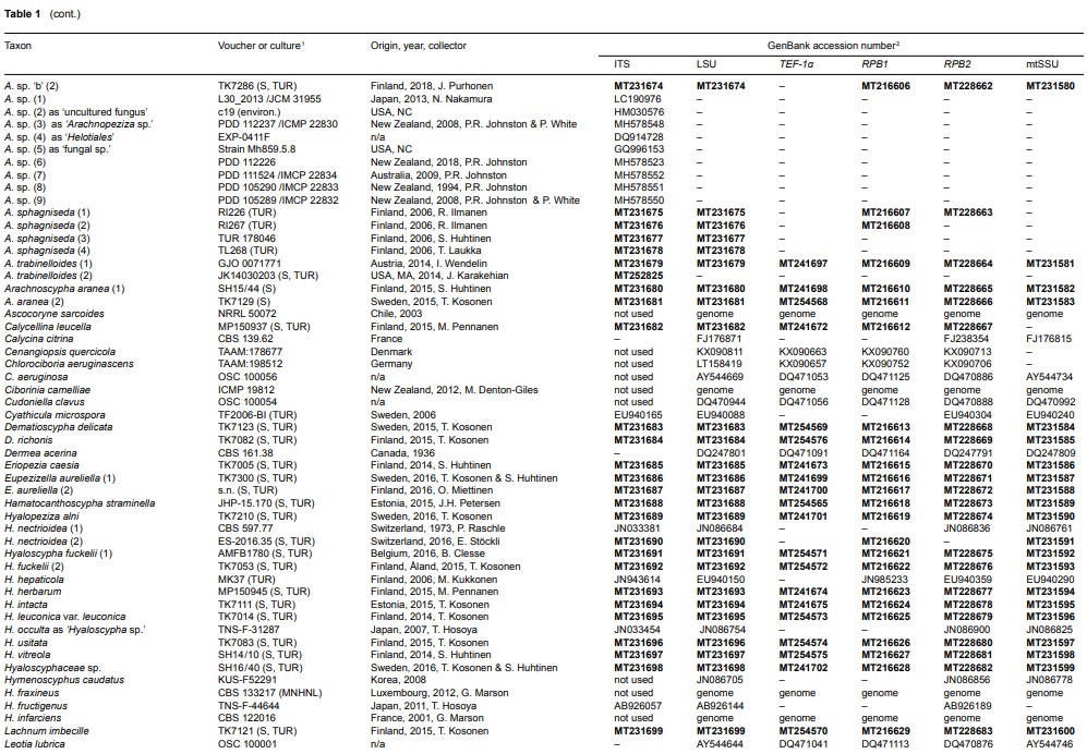
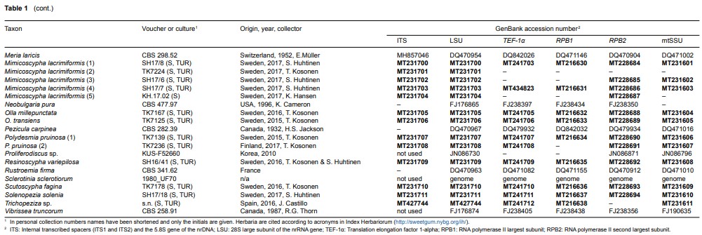
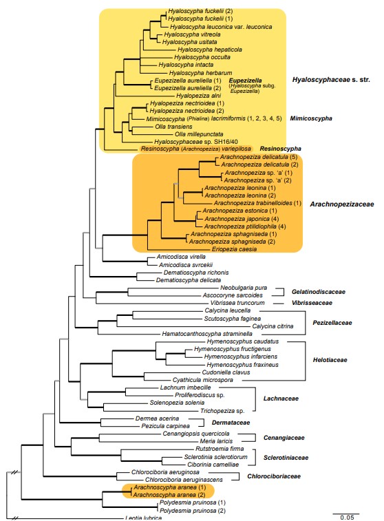
Fig. 2 Phylogenetic relationships of Arachnopezizaceae (orange square) and Hyaloscyphaceae (yellow square) among members of Helotiales based on Bayesian analyses of combined LSU, RPB1, RPB2, TEF-1α and mtSSU loci. Arachnoscypha aranea and Resinoscypha variepilosa (orange squares) were previously treated in Arachnopezizaceae. Thick black branches received both Bayesian posterior probabilities (PP) ≥ 0.95 and maximum likelihood bootstrap value (ML-BP) ≥ 75 %. Thick grey branches received support by either PP ≥ 0.95 or ML-BP ≥ 75 %. The number in parenthesis after a species name refers to an exact collection (see Table 1).
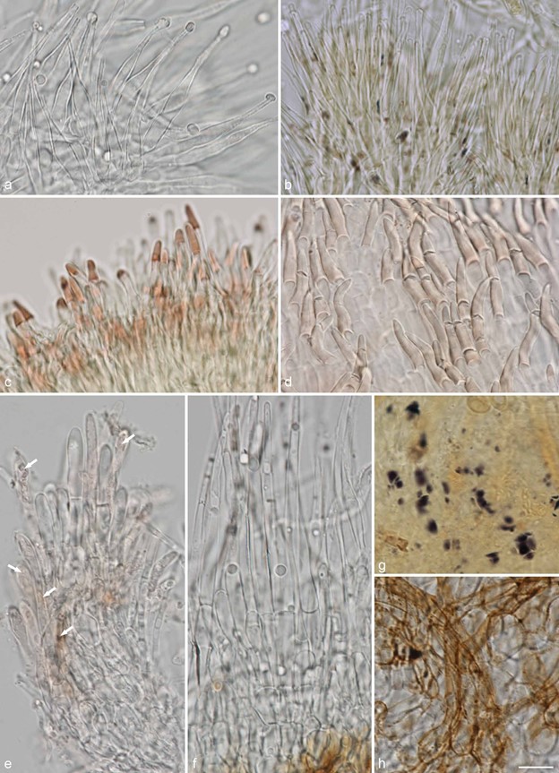
Fig. 14 Examples of hair shapes, inclusions and reactions, and of amyloid nodules in excipulum cells, in Hyaloscyphaceae. a. Thin-walled aseptate hairs, Hyaloscypha spiralis (in water); b. hairs showing amyloid and dextrinoid nodules, Eupezizella aureliella (in MLZ); c– d. dextrinoid glassy hairs in Olla (in MLZ): C.Olla transiens; d. Olla millepunctata; e. septate hairs showing resinous matter inside the hairs (arrows), Resinoscypha variepilosa (in water); f– h. Mimico- scypha lacrimiformis: f. septate hairs with refractive globules; g. purple nodules in ectal excipulum cells (in MLZ); h. bulbous basal cells of hairs with exudates (in water) (a: KH.16.02; b: T. Kosonen 7296; c: U. Söderholm 4829; d: T. Kosonen 7155; e: S. Huhtinen 16/41; f– h. KH.17.02). — Scale bars = 10 µm. — All from fresh material. — Photos: a, e– h. K. Hansen; b– d. T. Kosonen.
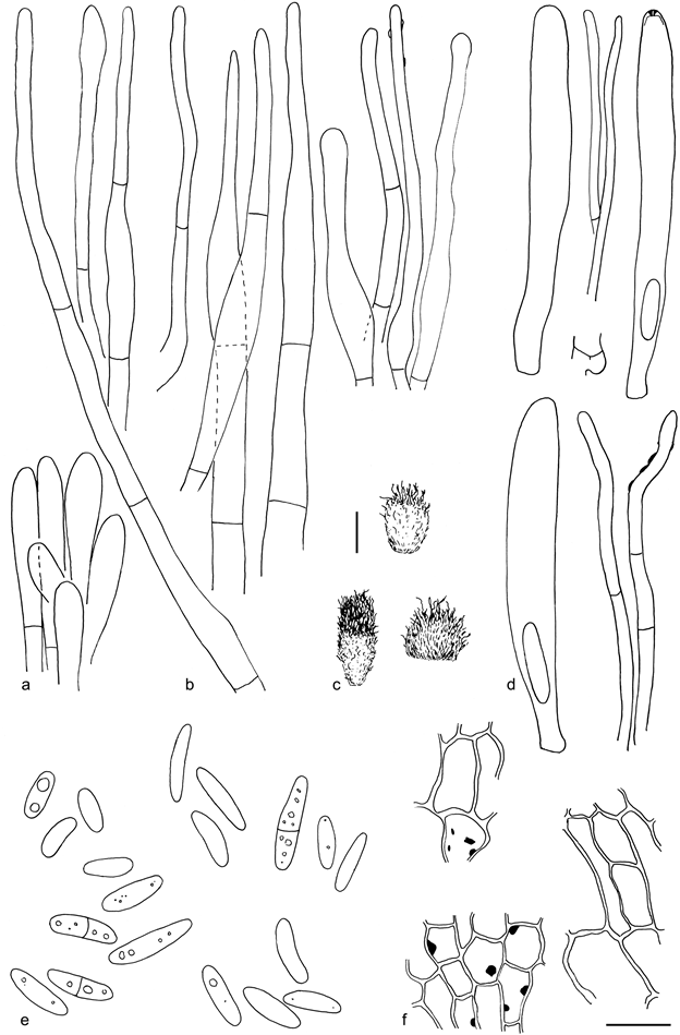
Fig. 15 Mimicoscypha lacrimiformis (holotype). a. Marginal end cells; b. marginal hairs in CR, the four on right in KOH; c. dry apothecia; d. asci and paraphyses;
e. spores; f. ectal excipulum in MLZ showing the amyloid nodules. — Scale bars: a– b, d– f = 10 µm, c = 100 µm. — Drawings: S. Huhtinen.
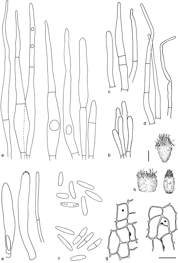
Fig. 16 Mimicoscypha lacrimiformis. a. Marginal hairs in CR showing several months old refractive globules; b. marginal end cells; c– d. marginal hairs in CR; e. asci and paraphysis; f. ascospores in CR; g. ectal excipulum in MLZ showing the amyloid nodules; h. dry apothecia (a, f: T. Kosonen 7224; b, d, g– h: S. Huhtinen 17/6; c, e: Huhtinen 17/8). — Scale bars: a– g = 10 µm, h = 100 µm. — Drawings: S. Huhtinen.
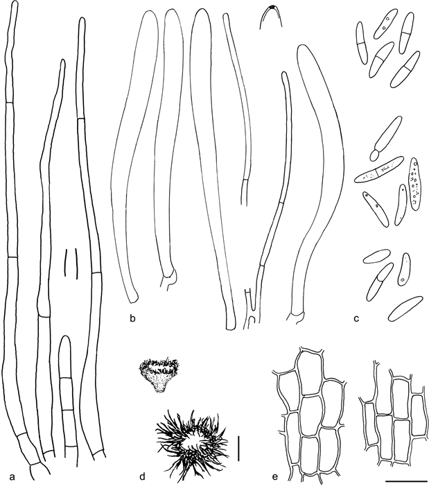
Fig. 17 Mimicoscypha mimica (holotype). a. Marginal hairs, detail showing maximal wall thickness in CR; b. asci and paraphyses; c. ascospores; d. dry apo- thecia; e. ectal excipulum in CB. — Scale bars: a– c, e = 10 µm, d = 100 µm. — Drawings: S. Huhtinen
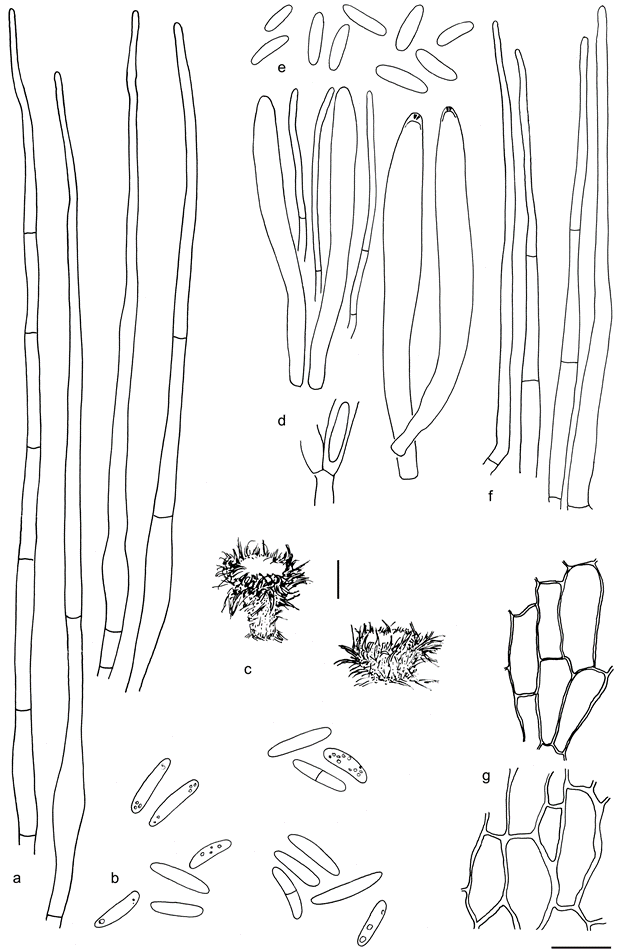
Fig. 18 Mimicoscypha paludosa. a. Marginal hairs in CR; b. ascospores; c. dry apothecia; d. asci and paraphyses; e. ascospores; f. marginal hairs; g. ectal excipulum in CR (a, g: PRM 817288; b– c, f (right): Clark 11 Sept. 1983; d– f (left): lectotype). — Scale bars: a– b, d– g = 10 µm, c = 100 µm. — Drawings: S. Huhtinen.
Species
