Mimicoscypha lacrimiformis (Hosoya) T. Kosonen, Huhtinen & K. Hansen, comb. nov.
MycoBank number: MB 835736; Index Fungorum number: IF 835736; Facesoffungi number: FoF 14071; Fig. 14f – h, 15, 16
Basionym. Phialina lacrimiformis Hosoya, Mycoscience 38: 181. 1997.
Holotype. JAPAN, Nagano Pref., Sanadacho, Daimyojin waterfall, on decaying wood, 24 May 1993, T. Hosoya (TNS-F-181594)
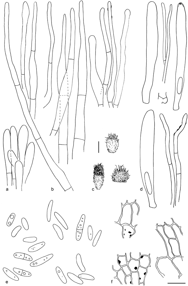
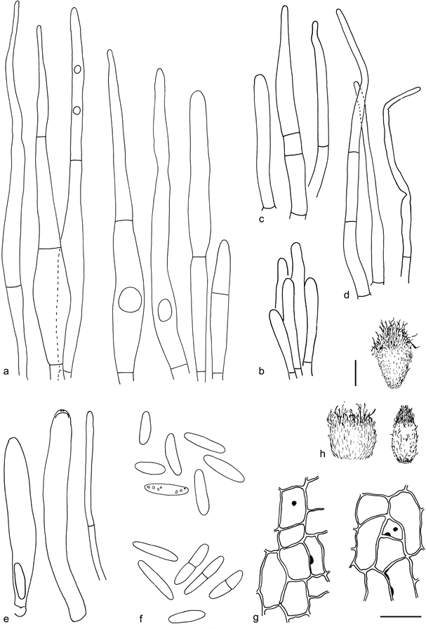
Apothecia 0.2 – 0.5 mm diam, white, disc-shaped and broadly attached, to shallowly cupulate, to shortly stipitate, prominently hairy at the margin. Ectal excipulum of textura prismatica to textura angularis, cell walls thickened, with abundant amyloid nodules in the basal excipulum cells, sometimes present also in cells closer to the margin, basal excipulum covered with brown, crustose resin, not changing in MLZ. Hairs up to 150 µm long, narrowly to broadly conical, straight, thin-walled and hence tardily reviving to their original shape, apex blunt or tapering to 1– 2 µm wide, close to base 4 – 8 µm wide, smooth, without apical solidifications, regularly with 1– 2 septa, rarely up to 6 –7 septa, occasionally with solitary refractive vacuoles inside, the amount varying between populations from prominent to lacking, external resinous matter gluing hairs together, present in water mounts or totally lacking, on lower flanks hairs occasionally completely covered in similar brown crustose resin as basal excipular cells. Asci 40 – 55 × 4 – 8 µm, cylindrical, 8-spored, apical pore MLZ+, arising from croziers. Ascospores 7.2 –12.6(–14) × 2.0 – 3.0 µm, mean 9.9 × 2.5 µm, Q = 3.2 – 4.8, mean Q = 4.0 (n = 28, from 3 populations), ellipsoid to oblong-ellipsoid to cylindrical, often slightly allantoid, aseptate, more rarely 1-septate, with or without refractive vacuoles when fresh in water. Paraphyses cylindrical, 1– 2 µm wide.
Specimens examined. SWEDEN, Östergötland, Ödeshög, Mörkahålkärrets Nature Reserve, on man-made coarse, coniferous wood chips, 26 Apr. 2017, S. Huhtinen 17/6 (S, TUR); same date, ecology and location, S. Huhtinen 17/7 (S, TUR); same date, ecology and location, S. Huhtinen 17/8 (S, TUR); same date, ecology and location, K. Hansen KH.17.02 (S, TUR); on a decayed trunk of Picea abies, T. Kosonen 7224 (S, TUR).
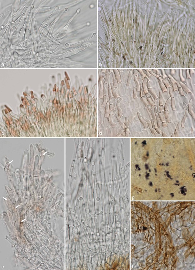
Notes — The absence of bright yellow pigment, typical for the genus Phialina (Huhtinen 1989), excludes its original placement by Hosoya (Hosoya & Otani 1997). In some populations from Sweden, there were clear refractive globules in the hairs when studied in fresh condition (e.g., Fig. 14f, 16a), whereas other populations, collected simultaneously from the same locality, lacked the character completely. The four collections sequenced by us had all identical ITS and LSU sequences. The RPB1, RPB2 and TEF-1α regions were in addition sequenced from two of these populations (S. Huhtinen 17/8, with refractive vacuoles and S. Huhtinen 17/7, without the vacuoles). All the sequences were identical between the two populations. The presence of amyloid nodules in the ectal excipulum (Fig. 14g, 15f, 16g) is a character common with Eupezizella and Resinoscypha. Swedish material of M. lacrimiformis shows marked variation in the amount of crustose, and brown resin present on basal excipulum cells and basal hairs (Fig. 14h). Even in the same collection, the apothecia vary from faintly coloured (as in the holotype) to basally dark brown. The species is new to Europe.

Fig. 14 Examples of hair shapes, inclusions and reactions, and of amyloid nodules in excipulum cells, in Hyaloscyphaceae. a. Thin-walled aseptate hairs, Hyaloscypha spiralis (in water); b. hairs showing amyloid and dextrinoid nodules, Eupezizella aureliella (in MLZ); c– d. dextrinoid glassy hairs in Olla (in MLZ): C.Olla transiens; d. Olla millepunctata; e. septate hairs showing resinous matter inside the hairs (arrows), Resinoscypha variepilosa (in water); f– h. Mimico- scypha lacrimiformis: f. septate hairs with refractive globules; g. purple nodules in ectal excipulum cells (in MLZ); h. bulbous basal cells of hairs with exudates (in water) (a: KH.16.02; b: T. Kosonen 7296; c: U. Söderholm 4829; d: T. Kosonen 7155; e: S. Huhtinen 16/41; f– h. KH.17.02). — Scale bars = 10 µm. — All from fresh material. — Photos: a, e– h. K. Hansen; b– d. T. Kosonen.

Fig. 15 Mimicoscypha lacrimiformis (holotype). a. Marginal end cells; b. marginal hairs in CR, the four on right in KOH; c. dry apothecia; d. asci and paraphyses; e. spores; f. ectal excipulum in MLZ showing the amyloid nodules. — Scale bars: a– b, d– f = 10 µm, c = 100 µm. — Drawings: S. Huhtinen.

Fig. 16 Mimicoscypha lacrimiformis. a. Marginal hairs in CR showing several months old refractive globules; b. marginal end cells; c– d. marginal hairs in CR; e. asci and paraphysis; f. ascospores in CR; g. ectal excipulum in MLZ showing the amyloid nodules; h. dry apothecia (a, f: T. Kosonen 7224; b, d, g– h: S. Huhtinen 17/6; c, e: Huhtinen 17/8). — Scale bars: a– g = 10 µm, h = 100 µm. — Drawings: S. Huhtinen.
