Trechispora subfissurata S.L. Liu, S.H. He & L.W. Zhou.
MycoBank number: MB 559899; Index Fungorum number: IF 559899; Facesoffungi number: FoF 12879;
Description
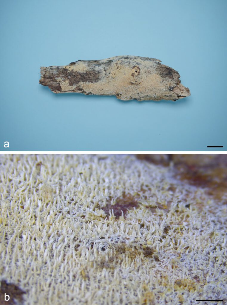
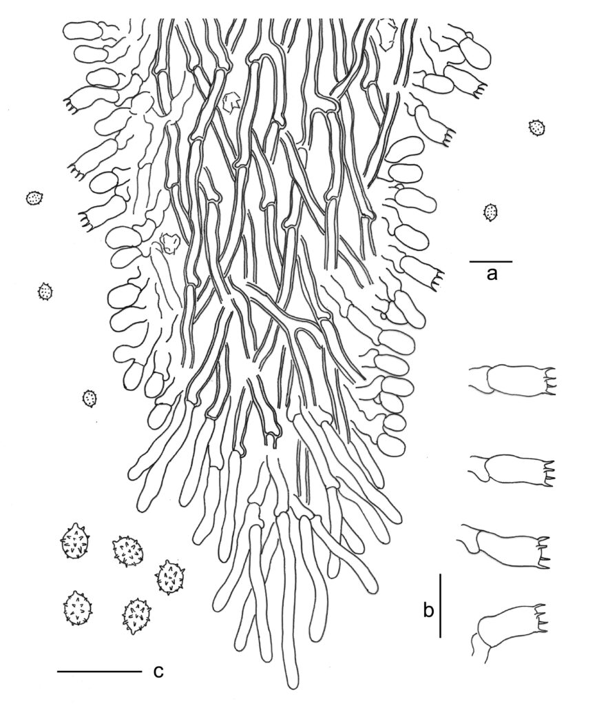
Basidiomes annual, resupinate, effused, thin, soft, fragile, easily separated from substrates, up to 8 cm long, 3 cm wide, 0.5 mm thick. Hymenophore odontioid to hydnoid with numerous aculei up to 0.4 mm long, white to cream when fresh, cream to buff-yellow when dry. Margin white to cream, fimbriate, up to 2 mm wide. Hyphal system monomitic; generative hyphae with clamp connections. Subicular generative hyphae hyaline, thick-walled, moderately branched and septate, subparallel, 2.5–4.5 µm in diam. Aculei composed of a central core of compact hyphae and subhymenial and hymenial layers; tramal generative hyphae distinct, hyaline, thick-walled, moderately branched, smooth, subparallel, 2.5–4 μm in diam. Cystidia absent. Crystals usually present, bipyramidic, aggregated. Basidia cylindrical with a slight median constriction, hyaline, thin-walled, with four sterigmata and a basal clamp connection, 11–15 × 4–5.5 µm; basidioles in shape similar to basidia, but slightly smaller. Basidiospores ellipsoid, hyaline, thin-walled, aculeate with spines that have a sharp apex, inamyloid, indextrinoid, acyanophilous, 2.8–3.5 × 2.5–3 µm, L = 3.1 µm, W = 2.8 µm, Q = 1.1 (n = 60/2).
Material examined: CHINA, Hainan, Baisha County, Yinggeling National Nature Reserve, on dead branch of living angiosperm, 9 June 2016, S.H. He, He 3907 (holotype in BJFC 022409).
Distribution: CHINA
Notes: Trechispora subfissurata is characterized by aculeate basidiospores with spines that have a sharp apex. This character makes T. subfissurata similar to T. echinospora and T. fissurata (Phookamsak et al. 2019, Zhao & Zhao 2021). However, T. echinospora differs in the absence of crystals in subiculum and trama, and globose basidiospores (Phookamsak et al. 2019), while T. fissurata differs in thicker basidiomes (up to 0.8 mm in thickness) with longer aculei (up to 0.9 mm in length; Zhao & Zhao 2021).

Fig. 1. Basidiomes of Trechispora subfissurata (He 3907, holotype). — Scale bars: a = 1 cm; b = 1 mm.

Fig. 2. Microscopic structures of Trechispora subfissurata (drawn from the holotype). a. Vertical section of basidiomes; b. Basidia; c. Basidiospores. — Scale bar = 10 μm.
