Trechispora sinensis S.L. Liu, L.W. Zhou & S.H. He.
MycoBank number: MB 559898; Index Fungorum number: IF 559898; Facesoffungi number: FoF 12878;
Description
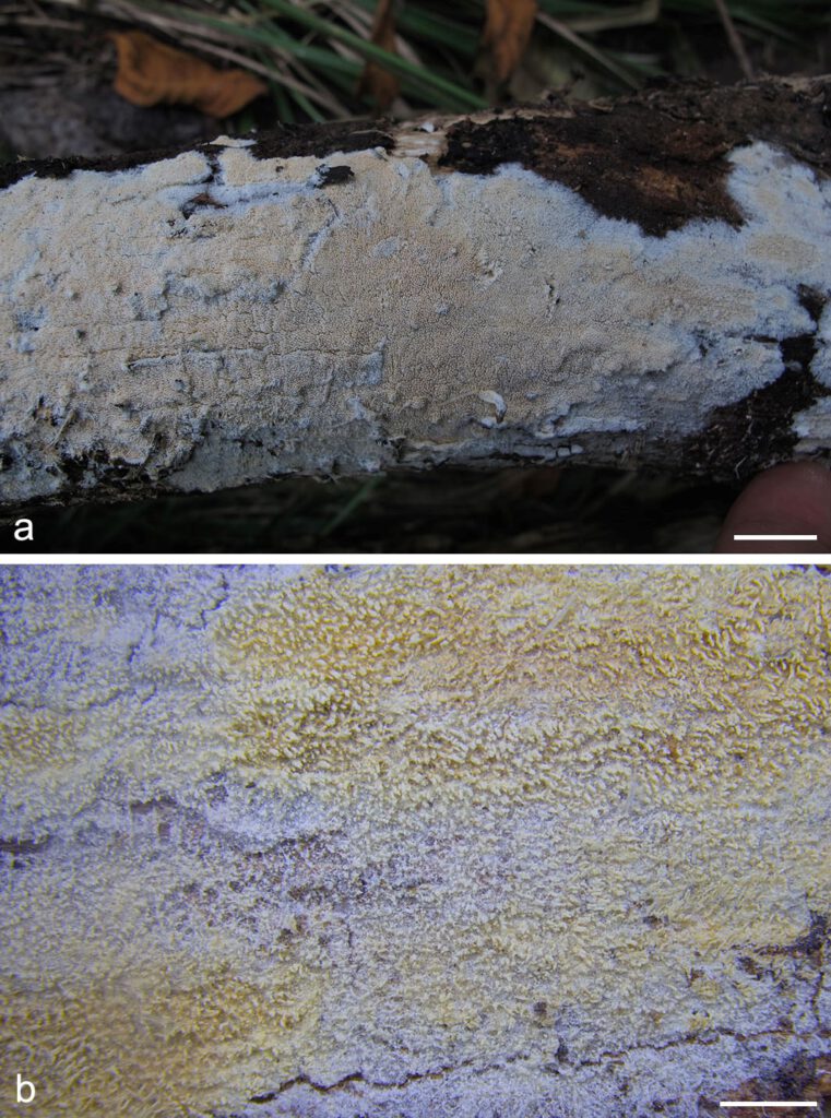
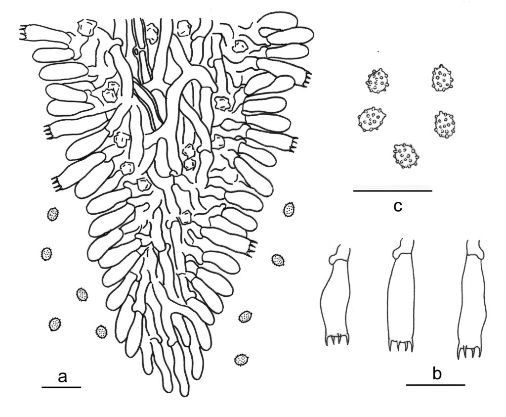
Basidiomes annual, resupinate, effused, thin, soft, fragile, easily separated from substrates, up to 12 cm long, 5 cm wide. Hymenophore odontioid with numerous small aculei, cream to straw-yellow when fresh, cinnamon-buff when dry. Margin white, fimbriate, up to 3 mm wide. Hyphal system monomitic; generative hyphae with clamp connections. Subicular hyphae hyaline, thin or thick-walled, moderately branched and septate, subparallel to interwoven, 2–3.5 µm in diam, ampullate septa up to 5 μm wide. Aculei composed of a central core of compact hyphae and subhymenial and hymenial layers; generative hyphae distinct, hyaline, thin or thick-walled, moderately branched, smooth, subparallel to interwoven, 2–3.5 μm in diam. Cystidia absent. Crystals usually present, bipyramidic, aggregated. Basidia cylindrical with a slight median constriction, thin-walled, with four sterigmata and a basal clamp connection, 11–15 × 3.8–5 μm; basidioles similar in shape to basidia, but smaller. Basidiospores broadly ellipsoid, hyaline, thin-walled, verrucose, inamyloid, indextrinoid, acyanophilous, 2.8–3.3 × (2.2–)2.5–2.9 μm, L = 3 μm, W = 2.7 μm, Q = 1.1–1.2 (n = 90/3).
Material examined: CHINA, Beijing, Mentougou, Donglingshan Scenic Spot, on fallen angiosperm branch, 4 Aug. 2018, L.W. Zhou, LWZ 20180804-19 (holotype in HMAS).
Distribution: CHINA
Notes: Trechispora sinensis has a wide distribution throughout China, including in multiple provincial regions, like Beijing, Chongqing, Guangdong, Guangxi, Guizhou, Hubei, Hunan, Jiangsu, Jiangxi, Jilin and Liaoning. Trechispora sinensis may be confused with T. chaibuxiensis and T. subsinensis; however, T. chaibuxiensis differs in the presence of hyphoid cystidia, while T. subsinensis differs in predominantly thick-walled tramal hyphae and aculeate basidiospores.

Fig. 1. Basidiomes of Trechispora sinensis (LWZ 20180804-19, holotype). — Scale bars: a = 1 cm; b = 2 mm.

Fig. 2. Microscopic structures of Trechispora sinensis (drawn from the holotype). a. Vertical section of basidiomes; b. Basidiospores. c. Basidia. — Scale bar = 10 μm.
