Trechispora subhymenocystis S.L. Liu, H.S. Yuan & L.W. Zhou.
MycoBank number: MB 559900; Index Fungorum number: IF 559900; Facesoffungi number: FoF 12880;
Description
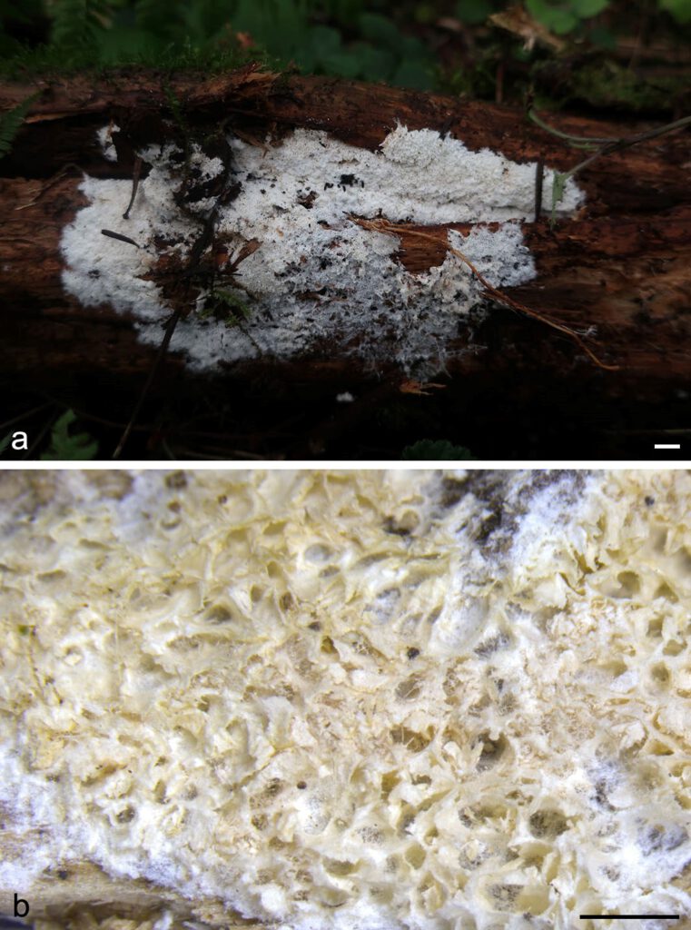
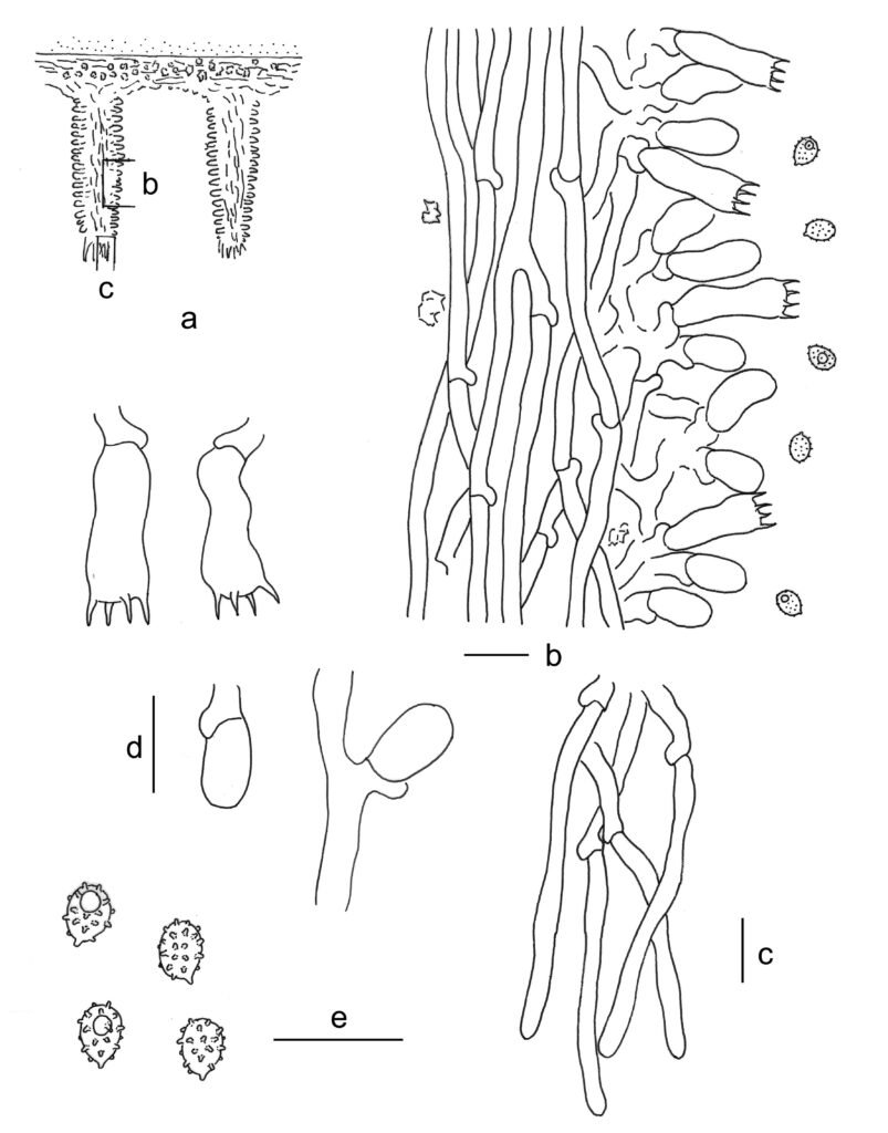
Basidiomes annual, resupinate, effused, soft, fragile, easily separated from substrates, soft when fresh, becoming soft corky when dry, up to 6 cm long, 4 cm wide. Pore surface white when fresh, buff, cinnamon-buff to yellowish brown when dry; pores round to angular, 3–4 per mm; dissepiments thin, entire. Subiculum white, soft corky, thin, about 0.1 mm thick. Tubes cinnamon-buff to yellowish brown, soft, up to 1.5 mm long. Margin white, fimbriate, rhizomorphic. Hyphal system monomitic; generative hyphae with clamp connections. Subicular generative hyphae hyaline, thin-walled, moderately branched and septate, subparallel, 2.5–5 µm in diam; crystals abundant, irregular. Generative hyphae in tubes hyaline, thin-walled, moderately branched and septate, more or less parallel, 2.5–4 µm in diam; crystals rare, irregular. Cystidia absent. Basidia cylindrical with a slight median constriction, hyaline, thin-walled, with four sterigmata and a basal clamp connection, 13–22 × 5–7 µm; basidioles in shape similar to basidia, but slightly smaller. Basidiospores ellipsoid, hyaline to yellowish, slightly thick-walled, aculeate with tubercles on spines, inamyloid, indextrinoid, acyanophilous, (3.5–)3.8–4.5(–4.8) × (2.8–)3–3.5(–3.9) µm, L = 4.1 µm, W = 3.1 µm, Q = 1.3 (n = 60/2).
Material examined: CHINA, Sichuan, Meigu County, Dafengding National Nature Reserve, on fallen trunk of Cryptomeria fortunei, 18 Aug. 2019, L.W. Zhou, LWZ 20190818-32b (holotype in HMAS).
Distribution: CHINA
Notes: The tuberculate ornamentations on spines of basidiospores, which were first reported in T. hymenocystis (Larsson 1994), make Trechispora subhymenocystis similar to T. araneosa, T. hondurensis, T. hymenocystis and T. minima. However, T. araneosa and T. minima differ in non-poroid hymenophores (Larsson 1995, 1996), T. hondurensis differs in smaller basidiospores (3.67–3.84 × 2.76–2.89 µm; Haelewaters et al. 2020), and T. hymenocystis differs in the presence of sphaerocysts in cords and the adjacent part of subiculum and larger basidiospores (4.5–5.5 × 3.5–4.5 µm; Larsson 1994). Trechispora subhymenocystis also resembles T. mollusca by poroid hymenophore, a monomitic hyphal system, and ellipsoid, aculeate basidiospores; however, T. mollusca differs in white to light ochraceous hymenophore, slightly thick-walled subicular hyphae and shorter basidiospores (2.5–4 µm in length; Liberta 1973, Larsson 1994, Bernicchia & Gorjón 2010).

Fig. 1. Basidiomes of Trechispora subhymenocystis (LWZ 20190818-32b, holotype). — Scale bars: a = 1 cm; b = 1 mm.

Fig. 2. Microscopic structures of Trechispora subhymenocystis (drawn from the holotype). a. Vertical section through basidiomes showing position of b and c; b. Section through a dissepiment edge; c. Section at the apex of a dissepiment; d. Basidia and basidioles; e. Basidiospores. — Scale bar = 10 μm.
