Sympodiorosea Q.V. Montoya & A. Rodrigues, gen. nov. (Fig. 6).
Index Fungorum number: MB 835147; Facesoffungi number: FoF
Etymology: “Sympodio” refers to the sympodial conidiog- enous cells, and “rosea” to the colony colour of the type species.
Diagnosis: Similar to Escovopsis and Luteomyces in the way it begins to grow, i.e., dense germination and form- ing stolon-like mycelia. However, Sympodiorosea differs from these genera and other known genera in the Hypo- creaceae by its holoblastic sympodial proliferous conid- iogenous cells.
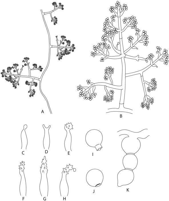
Description: Monophyletic genus in the Hypocreaceae. Colonies form inconspicuous to floccose, white, pale- beige, pink, brown aerial mycelia. Conidiophores formed on aerial mycelia (Fig. 6A), alternate or opposite, usually at right angles, with irregular branching conformation (Fig. 6A, B). Conidiogenous cells holoblastic, sympodial, proliferous (Fig. 6C–H), in pairs or in verticils on the apices of conidiophores and their branches, and soli- tary, alternate or opposite, on both the axes of the con- idiophore and their branches (Fig. 6B). Conidia formed solitary, globose to subglobose, smooth or rough (thick- walled), light-brown to dark-brown, with denticles or lesions like holes (Fig. 6I, J). Chlamydospores commonly formed (Fig. 6K).
Type species: Sympodiorosea kreiselii (L.A. Meirelles et al.) Q.V. Montoya & A. Rodrigues 2021.
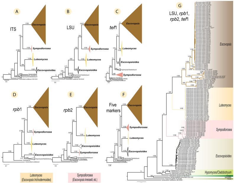
Notes: – Sympodiorosea is phylogenetically placed in the Hypocreaceae near the genera Luteomyces and Escovop- sioides (Fig. 3G). However, Sympodiorosea grows slower and has different colony colour (pink) than Luteomy- ces (yellow) and Escovopsioides (white). Sympodiorosea forms more branched conidiophores than Luteomyces and Escovopsioides.

Fig. 3 Phylogenetic placement of Escovopsis, Sympodiorosea and Luteomyces within the Hypocreaceae. Phylogenies shown were inferred using Bayesian Inference (BI) and are separated for each region (molecular marker): (A) ITS, (B) LSU, (C) tef1, (D) rpb1, and (E) rpb2. The remaining two trees are based on concatenated datasets of (F) all five markers, and (G) four of the five markers (LSU, rpb1, rpb2, and tef1). Numbers on branches indicate BI posterior probabilities (PP) and Maximum Likelihood bootstrap support values (MLB), respectively. Hyphens (–) indicate MLB < 70%. Lecanicillium antillanum CBS 350.85 was used as the outgroup. ET indicates ex-type cultures. See Table S1 for all strains and their associated metadata used to infer these phylogenetic trees

Fig. 6 Illustration of the microscopic structures of Sympodiorosea kreiselii (syn. Escovopsis kreiselii). A Pattern of disposition of conidiophores on aerial mycelium. B A conidiophore. C–H Shapes of the sympodial conidiogenous cells. I Conidia with denticle (detached from the conidiogenous cell), J Conidia with a hole (when the denticle remains on the conidiogenous cell). K Chlamydospores in chain
Species
