Stachybotrys renispora P.C. Misra, Mycotaxon 4: 161 (1976), Index Fungorum Number: 626938
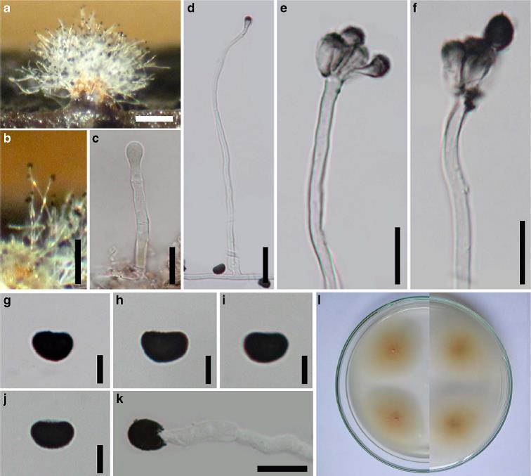
Saprobic on dead twig of T. grandis. Sexual morph: Undetermined. Asexual morph: Colonies superficial, gregarious, white, erumpent, bouquet-like, velvety, developing on wood after incubation in a moist chamber for 7 days. Conidiophores (48–)126–131(183–) μm long, 2–4μm wide (x = 102×3μm, n = 15), erect or flexuous, fasciculate, hyaline, smooth, dark grey and verrucose near the tip, 1–2-septate, branched. Conidiogenous cells up to 4 on each conidiophore 8–10× 3–4 (x = 9×4μm, n = 10), monophialidic, with an indistinct collarette, discrete, pigmented, darkened at the apex, clavate, ellipsoidal to broadly fusiform, smooth. Conidia 6– 9×4–6μm (x = 8×5μm, n = 20), reniform, fusiform, rounded at both ends, slightly curved, blackish brown or black, numerous, smooth or verrucose, aseptate.
Culture characteristics: Conidia germinating on PDA after 24 h. Germ tubes produced from germ pores. Colonies growing on MEA reaching 19–21 mm diam. after 3 weeks in the dark at 25 °C, (x = 18.9, n = 5), crenated, low convex, cottony, medium dense, aerial, grey (4D1) at the old mycelium plugs and white (4A1) at the edge from above, greyish yellowish (4B3) from below.
Habitat: Known to inhabit seed of Phlox drummondii (Misra 1976) and dead twig of T. grandis (Doilom et al. 2017).
Known distribution: India (Misra 1976) and Thailand (Doilom et al. 2017).
Material examined: THAILAND, Chiang Mai Province, Chiang Dao District, on dead twig of T. grandis, (incubation
in moist chamber after 7 days), 22 July 2012, M. Doilom & E. Wongsakul, MFLU 15–3424, living culture MFLUCC 12– 0553, MKT 069, ICMP 21163, GenBank Accession No: ITS: KU144929, SSU: KU712477.
Notes: The conidia of Stachybotrys renispora in this study are reniform or kidney-shaped and according to the recent key of Stachybotrys species (Wang et al. 2015), ten species produce reniform conidia. These include St. nephrodes McKenzie, St. nephrospora Hansf., St. oenanthes M.B. Ellis, St. proliferata K.G. Karand. et al., St. reniformis Tubaki, St. renispora P.C. Misra, St. renisporoides K.G. Karand. et al., St. reniverrucosa Whitton et al., St. sinuatophora Matsush. and St. subreniformis Q.R. Li & Y.L. Jiang. The conidial shape in the present collections is rather similar to St. renispora, St. reniformis and St. subreniformis. However the conidiophores of St. reniformis are unbranched while the material in this study, and those of St. renispora and St. subreniformis are branched. The present collections also resemble St. renispora (type) by having verrucose conidiophores while St. subreniformis has smooth conidiophores. Conidial size in this study 5.9–8.5 × 4.5–5.3 (x = 7.3×5μm) is also similar to St. renispora 5.2–7× 3.5–5.2 (x not reported) (Misra 1976) although they were observed on different substrates (on dead twig of T. grandis versus on MEA). The MegaBLAST search of NCBIs GenBank nucleotide database results of the ITS sequence data of isolate MFLUCC 12–0553 (accession number KU144929) showed Identities= 486/488 (99 %) and 486/489 (99 %) similarity to St. nephrospora (Stanep1907, GenBank KF626486 and Stanep2207, KF626488). However, these sequences are unpublished and no ex-type sequence data of St. renispora is available in GenBank (Wang et al. 2015). Based on morphological features of the conidia size and shape as well as conidiophore texture, the collections in the present study are considered to be St. renispora.
FIG: Stachybotrys renispora (MFLU15–3424). a, b Colonies on host surface after incubation in a moist chamber for 7 days. c Developing conidiogenous cell on conidiophore. d Conidiophore with conidiogenous cells. e Close-up conidiogenous cells attached to conidiophore. f Close-up conidium attached to conidiogenous cell and conidiophores. g–j Reniform conidia. k Germinated conidium. l Colonies on PDA after 4 days from single spore isolation. Scale bars: a, b = 100 μm, c, e, f, k = 10μm, d = 20μm, g–j = 5μm